Show PDB file:   
         Plain Text   HTML   (compressed file size)
QuickSearch:   
by PDB,NDB,UniProt,PROSITE Code or Search Term(s)  
(-)NMR Structure - model 1
(-)NMR Structure - model 1, sites
(-)NMR Structure - all models
collapse expand < >
Image NMR Structure - model 1
NMR Structure - model 1  (Jmol Viewer)
Image NMR Structure - model 1, sites
NMR Structure - model 1, sites  (Jmol Viewer)
Image NMR Structure - all models
NMR Structure - all models  (Jmol Viewer)

(-) Description

Title :  STRUCTURE OF CALCIUM-SATURATED CARDIAC TROPONIN C, NMR, 1 STRUCTURE
 
Authors :  S. K. Sia, M. X. Li, L. Spyracopoulos, S. M. Gagne, W. Liu, J. A. Putkey, B. D. Sykes
Date :  14 May 97  (Deposition) - 20 May 98  (Release) - 19 May 09  (Revision)
Method :  SOLUTION NMR
Resolution :  NOT APPLICABLE
Chains :  NMR Structure  :  A
Keywords :  Cardiac, Muscle Protein, Regulatory, Calcium Binding (Keyword Search: [Gene Ontology, PubMed, Web (Google))
 
Reference :  S. K. Sia, M. X. Li, L. Spyracopoulos, S. M. Gagne, W. Liu, J. A. Putkey, B. D. Sykes
Structure Of Cardiac Muscle Troponin C Unexpectedly Reveals A Closed Regulatory Domain.
J. Biol. Chem. V. 272 18216 1997
PubMed-ID: 9218458  |  Reference-DOI: 10.1074/JBC.272.29.18216
(for further references see the PDB file header)

(-) Compounds

Molecule 1 - TROPONIN C
    Cellular LocationTHIN FILAMENT
    Cell LineBL21
    ChainsA
    EngineeredYES
    Expression SystemESCHERICHIA COLI
    Expression System GeneCTNC(A-CYS)
    Expression System PlasmidPET-23D
    Expression System StrainBL21 (DE3) PLYSS
    Expression System Taxid562
    GeneCTNC(A-CYS)
    MutationYES
    OrganHEART
    Organism CommonCHICKEN
    Organism ScientificGALLUS GALLUS
    Organism Taxid9031
    Other DetailsCARDIAC TROPONIN C, CALCIUM IONS BOUND AT SITES II, III AND IV
    SynonymCTNC
    TissueMUSCLE

 Structural Features

(-) Chains, Units

  
NMR Structure 

Summary Information (see also Sequences/Alignments below)

(-) Ligands, Modified Residues, Ions  (1, 3)

NMR Structure (1, 3)
No.NameCountTypeFull Name
1CA3Ligand/IonCALCIUM ION

(-) Sites  (3, 3)

NMR Structure (3, 3)
No.NameEvidenceResiduesDescription
1AC1SOFTWAREASP A:65 , ASP A:67 , SER A:69 , THR A:71 , GLU A:76BINDING SITE FOR RESIDUE CA A 162
2AC2SOFTWAREASP A:105 , ASN A:107 , ASP A:109 , TYR A:111 , ILE A:112 , GLU A:116BINDING SITE FOR RESIDUE CA A 163
3AC3SOFTWAREASP A:141 , ASN A:143 , ASP A:145 , ARG A:147 , GLU A:152BINDING SITE FOR RESIDUE CA A 164

(-) SS Bonds  (0, 0)

(no "SS Bond" information available for 1AJ4)

(-) Cis Peptide Bonds  (0, 0)

(no "Cis Peptide Bond" information available for 1AJ4)

 Sequence-Structure Mapping

(-) SAPs(SNPs)/Variants  (0, 0)

(no "SAP(SNP)/Variant" information available for 1AJ4)

(-) PROSITE Motifs  (2, 7)

NMR Structure (2, 7)
 PROSITEUniProtKBPDB
No.IDACDescriptionIDLocationCountLocation
1EF_HAND_2PS50222 EF-hand calcium-binding domain profile.TNNC1_CHICK33-51
52-87
92-127
128-161
  4A:33-51
A:52-87
A:92-127
A:128-161
2EF_HAND_1PS00018 EF-hand calcium-binding domain.TNNC1_CHICK65-77
105-117
141-153
  3A:65-77
A:105-117
A:141-153

(-) Exons   (6, 6)

NMR Structure (6, 6)
 ENSEMBLUniProtKBPDB
No.Transcript IDExonExon IDGenome LocationLengthIDLocationLengthCountLocationLength
1.1ENSGALT000000022141ENSGALE00000013329chr12:545708-54566049TNNC1_CHICK1-881A:2-87
1.2ENSGALT000000022142ENSGALE00000013327chr12:542533-54250331TNNC1_CHICK9-19111A:9-1911
1.3ENSGALT000000022143ENSGALE00000013330chr12:542326-542180147TNNC1_CHICK19-68501A:19-6850
1.4ENSGALT000000022144ENSGALE00000013326chr12:541358-541244115TNNC1_CHICK68-106391A:68-10639
1.5ENSGALT000000022145ENSGALE00000013328chr12:541140-541004137TNNC1_CHICK106-152471A:106-15247
1.6ENSGALT000000022146ENSGALE00000013325chr12:540437-540264174TNNC1_CHICK152-161101A:152-16110

(-) Sequences/Alignments

NMR Structure
   Reformat: Number of residues per line =  ('0' or empty: single-line sequence representation)
  Number of residues per labelling interval =   
  UniProt sequence: complete  aligned part    
   Show mapping: SCOP domains CATH domains Pfam domains Secondary structure (by author)
SAPs(SNPs) PROSITE motifs Exons
(details for a mapped element are shown in a popup box when the mouse pointer rests over it)
Chain A from PDB  Type:PROTEIN  Length:160
 aligned with TNNC1_CHICK | P09860 from UniProtKB/Swiss-Prot  Length:161

    Alignment length:160
                                    11        21        31        41        51        61        71        81        91       101       111       121       131       141       151       161
          TNNC1_CHICK     2 DDIYKAAVEQLTEEQKNEFKAAFDIFVLGAEDGCISTKELGKVMRMLGQNPTPEELQEMIDEVDEDGSGTVDFDEFLVMMVRCMKDDSKGKTEEELSDLFRMFDKNADGYIDLEELKIMLQATGETITEDDIEELMKDGDKNNDGRIDYDEFLEFMKGVE 161
               SCOP domains d1aj4a_ A: Troponin C                                                                                                                                            SCOP domains
               CATH domains 1aj4A00 A:2-161 EF-hand                                                                                                                                          CATH domains
               Pfam domains ---------------------------------------------------------------------------------------------------------------------------------------------------------------- Pfam domains
         Sec.struct. author ....hhhhh...hhhhhhhhhhhhhhh......eeehhhhhhhhhhh.....hhhhhhhhhhh......eeehhhhhhhhhh...........hhhhhhhhh.......eeehhhhhhhhhh......hhhhhhhhhhh......eeehhhhhhhhh... Sec.struct. author
                 SAPs(SNPs) ---------------------------------------------------------------------------------------------------------------------------------------------------------------- SAPs(SNPs)
                PROSITE (1) -------------------------------EF_HAND_2          EF_HAND_2  PDB: A:52-87             ----EF_HAND_2  PDB: A:92-127            EF_HAND_2  PDB: A:128-161          PROSITE (1)
                PROSITE (2) ---------------------------------------------------------------EF_HAND_1    ---------------------------EF_HAND_1    -----------------------EF_HAND_1    -------- PROSITE (2)
           Transcript 1 (1) 1.1    Exon 1.2   ------------------------------------------------Exon 1.4  PDB: A:68-106 UniProt: 68-106---------------------------------------------Exon 1.6   Transcript 1 (1)
           Transcript 1 (2) -----------------Exon 1.3  PDB: A:19-68 UniProt: 19-68             -------------------------------------Exon 1.5  PDB: A:106-152 UniProt: 106-152      --------- Transcript 1 (2)
                 1aj4 A   2 ADIYKAAVEQLTEEQKNEFKAAFDIFVLGAEDGSISTKELGKVMRMLGQNPTPEELQEMIDEVDEDGSGTVDFDEFLVMMVRSMKDDSKGKTEEELSDLFRMFDKNADGYIDLEELKIMLQATGETITEDDIEELMKDGDKNNDGRIDYDEFLEFMKGVE 161
                                    11        21        31        41        51        61        71        81        91       101       111       121       131       141       151       161

   Legend:   → Mismatch (orange background)
  - → Gap (green background, '-', border residues have a numbering label)
    → Modified Residue (blue background, lower-case, 'x' indicates undefined single-letter code, labelled with number + name)
  x → Chemical Group (purple background, 'x', labelled with number + name, e.g. ACE or NH2)
  extra numbering lines below/above indicate numbering irregularities and modified residue names etc., number ends below/above '|'

 Classification and Annotation

(-) SCOP Domains  (1, 1)

NMR Structure

(-) CATH Domains  (1, 1)

NMR Structure

(-) Pfam Domains  (0, 0)

(no "Pfam Domain" information available for 1AJ4)

(-) Gene Ontology  (17, 17)

NMR Structure(hide GO term definitions)
Chain A   (TNNC1_CHICK | P09860)
molecular function
    GO:0051015    actin filament binding    Interacting selectively and non-covalently with an actin filament, also known as F-actin, a helical filamentous polymer of globular G-actin subunits.
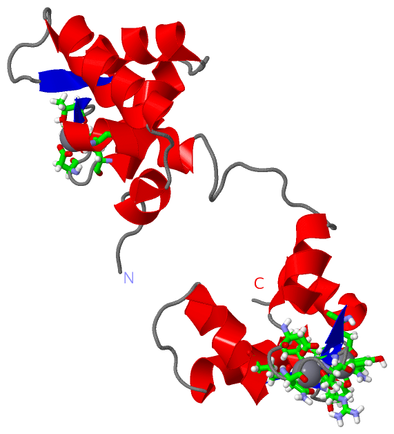
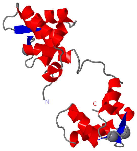
    GO:0005509    calcium ion binding    Interacting selectively and non-covalently with calcium ions (Ca2+).
    GO:0048306    calcium-dependent protein binding    Interacting selectively and non-covalently with any protein or protein complex (a complex of two or more proteins that may include other nonprotein molecules), in the presence of calcium.
    GO:0046872    metal ion binding    Interacting selectively and non-covalently with any metal ion.
    GO:0042803    protein homodimerization activity    Interacting selectively and non-covalently with an identical protein to form a homodimer.
    GO:0031013    troponin I binding    Interacting selectively and non-covalently with troponin I, the inhibitory subunit of the troponin complex.
    GO:0031014    troponin T binding    Interacting selectively and non-covalently with troponin T, the tropomyosin-binding subunit of the troponin complex.
biological process
    GO:0060048    cardiac muscle contraction    Muscle contraction of cardiac muscle tissue.
    GO:0043462    regulation of ATPase activity    Any process that modulates the rate of ATP hydrolysis by an ATPase.
    GO:0006937    regulation of muscle contraction    Any process that modulates the frequency, rate or extent of muscle contraction.
    GO:0032972    regulation of muscle filament sliding speed    Any process that modulates the velocity of muscle filament sliding.
    GO:0014883    transition between fast and slow fiber    The process of conversion of fast-contracting muscle fibers to a slower character. This may involve slowing of contractile rate, slow myosin gene induction, increase in oxidative metabolic properties, altered electrophysiology and altered innervation. This process also regulates skeletal muscle adapatation.
    GO:0055010    ventricular cardiac muscle tissue morphogenesis    The process in which the anatomical structures of cardiac ventricle muscle is generated and organized.
cellular component
    GO:0015629    actin cytoskeleton    The part of the cytoskeleton (the internal framework of a cell) composed of actin and associated proteins. Includes actin cytoskeleton-associated complexes.
    GO:0005739    mitochondrion    A semiautonomous, self replicating organelle that occurs in varying numbers, shapes, and sizes in the cytoplasm of virtually all eukaryotic cells. It is notably the site of tissue respiration.
    GO:0005654    nucleoplasm    That part of the nuclear content other than the chromosomes or the nucleolus.
    GO:0005861    troponin complex    A complex of accessory proteins (typically troponin T, troponin I and troponin C) found associated with actin in muscle thin filaments; involved in calcium regulation of muscle contraction.

 Visualization

(-) Interactive Views

NMR Structure
  Complete Structure
    Jena3D(integrated viewing of ligand, site, SAP, PROSITE, SCOP information)
    WebMol | AstexViewer[tm]@PDBe
(Java Applets, require no local installation except for Java; loading may be slow)
    STRAP
(Java WebStart application, automatic local installation, requires Java; full application with system access!)
    RasMol
(require local installation)
    Molscript (VRML)
(requires installation of a VRML viewer; select preferred view via VRML and generate a mono or stereo PDF format file)
 
  Ligands, Modified Residues, Ions
    CA  [ RasMol | Jena3D ]  +environment [ RasMol | Jena3D ]
 
  Sites
    AC1  [ RasMol ]  +environment [ RasMol ]
    AC2  [ RasMol ]  +environment [ RasMol ]
    AC3  [ RasMol ]  +environment [ RasMol ]
 
  Cis Peptide Bonds
(no "Cis Peptide Bonds" information available for 1aj4)
 

(-) Still Images

Jmol
  protein: cartoon or spacefill or dots and stick; nucleic acid: cartoon and stick; ligands: spacefill; active site: stick
Molscript
  protein, nucleic acid: cartoon; ligands: spacefill; active site: ball and stick

 Databases and Analysis Tools

(-) Databases

Access by PDB/NDB ID
  1aj4
    Family and Domain InformationProDom | SYSTERS
    General Structural InformationGlycoscienceDB | MMDB | NDB | OCA | PDB | PDBe | PDBj | PDBsum | PDBWiki | PQS | PROTEOPEDIA
    Orientation in MembranesOPM
    Protein SurfaceSURFACE
    Secondary StructureDSSP (structure derived) | HSSP (homology derived)
    Structural GenomicsGeneCensus
    Structural NeighboursCE | VAST
    Structure ClassificationCATH | Dali | SCOP
    Validation and Original DataBMRB Data View | BMRB Restraints Grid | EDS | PROCHECK | RECOORD | WHAT_CHECK
 
Access by UniProt ID/Accession number
  TNNC1_CHICK | P09860
    Comparative Protein Structure ModelsModBase
    Genomic InformationEnsembl
    Protein-protein InteractionDIP
    Sequence, Family and Domain InformationInterPro | Pfam | SMART | UniProtKB/SwissProt
 
Access by Enzyme Classificator   (EC Number)
  (no 'Enzyme Classificator' available)
    General Enzyme InformationBRENDA | EC-PDB | Enzyme | IntEnz
    PathwayKEGG | MetaCyc
 
Access by Disease Identifier   (MIM ID)
  (no 'MIM ID' available)
    Disease InformationOMIM
 
Access by GenAge ID
  (no 'GenAge ID' available)
    Age Related InformationGenAge

(-) Analysis Tools

Access by PDB/NDB ID
    Domain InformationXDom
    Interatomic Contacts of Structural UnitsCSU
    Ligand-protein ContactsLPC
    Protein CavitiescastP
    Sequence and Secondary StructurePDBCartoon
    Structure AlignmentSTRAP(Java WebStart application, automatic local installation, requires Java; full application with system access!)
    Structure and Sequence BrowserSTING
 
Access by UniProt ID/Accession number
  TNNC1_CHICK | P09860
    Protein Disorder PredictionDisEMBL | FoldIndex | GLOBPLOT (for more information see DisProt)

 Related Entries

(-) Entries Sharing at Least One Protein Chain (UniProt ID)

UniProtKB/Swiss-Prot
        TNNC1_CHICK | P098601dtl 1fi5 1la0 1sbj 1scv 2ctn 3ctn

(-) Related Entries Specified in the PDB File

(no "Related Entries Specified in the PDB File" available for 1AJ4)