| molecular function |
|---|
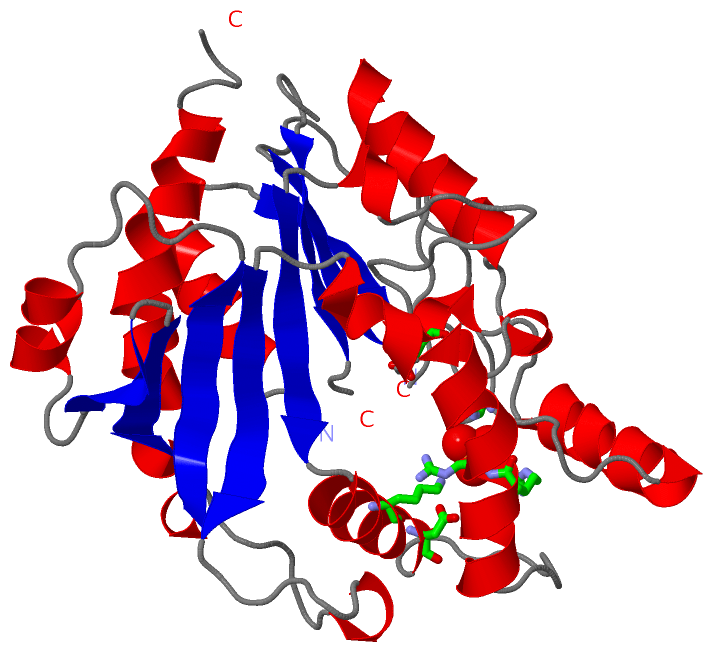
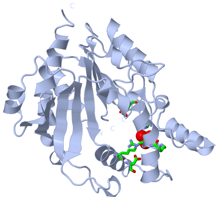
| | GO:0005524 | | ATP binding | | Interacting selectively and non-covalently with ATP, adenosine 5'-triphosphate, a universally important coenzyme and enzyme regulator. |
| | GO:0016301 | | kinase activity | | Catalysis of the transfer of a phosphate group, usually from ATP, to a substrate molecule. |
| | GO:0000166 | | nucleotide binding | | Interacting selectively and non-covalently with a nucleotide, any compound consisting of a nucleoside that is esterified with (ortho)phosphate or an oligophosphate at any hydroxyl group on the ribose or deoxyribose. |
| | GO:0008974 | | phosphoribulokinase activity | | Catalysis of the reaction: D-ribulose 5-phosphate + ATP = D-ribulose 1,5-bisphosphate + ADP + 2 H(+). |
| | GO:0016740 | | transferase activity | | Catalysis of the transfer of a group, e.g. a methyl group, glycosyl group, acyl group, phosphorus-containing, or other groups, from one compound (generally regarded as the donor) to another compound (generally regarded as the acceptor). Transferase is the systematic name for any enzyme of EC class 2. |
| biological process |
|---|
| | GO:0005975 | | carbohydrate metabolic process | | The chemical reactions and pathways involving carbohydrates, any of a group of organic compounds based of the general formula Cx(H2O)y. Includes the formation of carbohydrate derivatives by the addition of a carbohydrate residue to another molecule. |
| | GO:0008152 | | metabolic process | | The chemical reactions and pathways, including anabolism and catabolism, by which living organisms transform chemical substances. Metabolic processes typically transform small molecules, but also include macromolecular processes such as DNA repair and replication, and protein synthesis and degradation. |
| | GO:0016310 | | phosphorylation | | The process of introducing a phosphate group into a molecule, usually with the formation of a phosphoric ester, a phosphoric anhydride or a phosphoric amide. |
| | GO:0015979 | | photosynthesis | | The synthesis by organisms of organic chemical compounds, especially carbohydrates, from carbon dioxide (CO2) using energy obtained from light rather than from the oxidation of chemical compounds. |
| | GO:0019253 | | reductive pentose-phosphate cycle | | The fixation of carbon dioxide (CO2) as glucose in the chloroplasts of C3 plants; uses ATP and NADPH formed in the light reactions of photosynthesis; carbon dioxide reacts with ribulose 1,5-bisphosphate (catalyzed by the function of ribulose-bisphosphate carboxylase) to yield two molecules of 3-phosphoglycerate; these are then phosphorylated by ATP to 1,3-bisphosphateglyceraldehyde which, in turn, is then reduced by NADPH to glyceraldehyde 3-phosphate. The glyceraldehyde 3-phosphate is converted to fructose 5-phosphate and ribulose 5-phosphate by aldolase and other enzymes; the ribulose 5-phosphate is phosphorylated by ATP to ribulose 1,5-bisphosphate. |
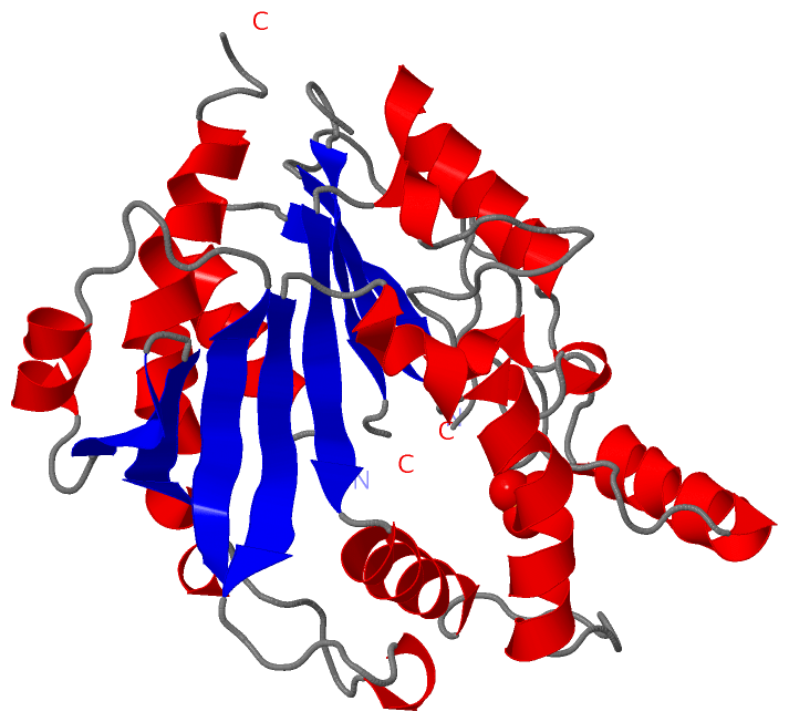
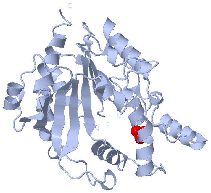
 Description