|
|
|
|
 Description
|
|
Compounds
|
||||||||||||||||||||||||||||||||||||||||||||||||||
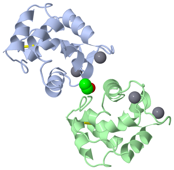
Chains, Units
Summary Information (see also Sequences/Alignments below) |
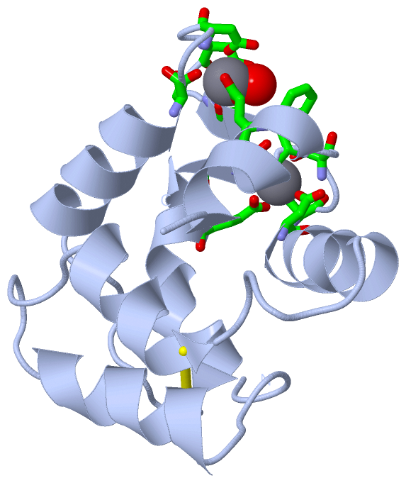
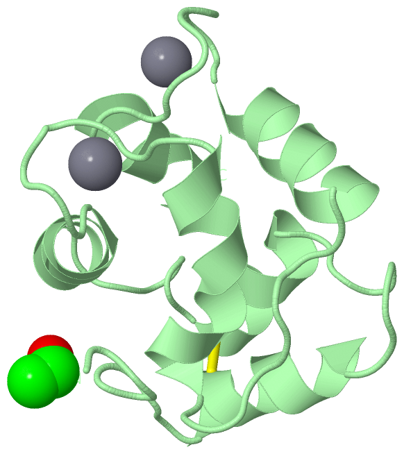
Ligands, Modified Residues, Ions (2, 5)| Asymmetric Unit (2, 5) Biological Unit 1 (0, 0) Biological Unit 2 (1, 1) |
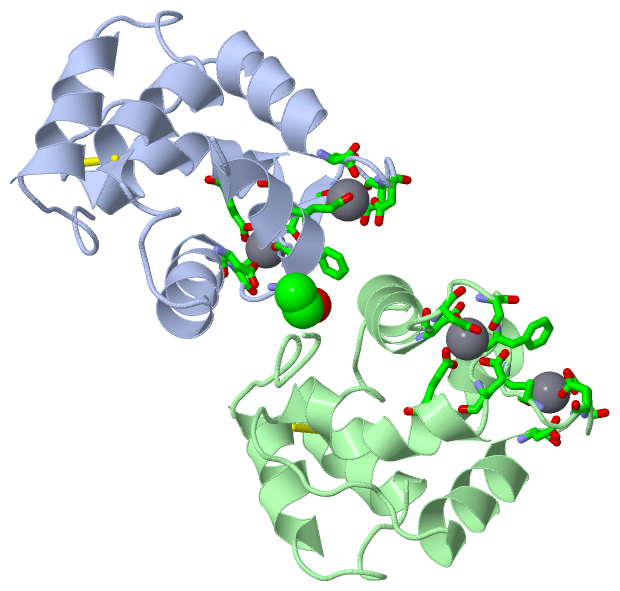
Sites (8, 8)
Asymmetric Unit (8, 8)
|
SS Bonds (2, 2)
Asymmetric Unit
|
||||||||||||
Cis Peptide Bonds (0, 0)| (no "Cis Peptide Bond" information available for 1A75) |
SAPs(SNPs)/Variants (0, 0)| (no "SAP(SNP)/Variant" information available for 1A75) |
PROSITE Motifs (2, 8)
Asymmetric Unit (2, 8)
|
||||||||||||||||||||||||||||||||||||||||||||||||||||||||||||||||||||||||||||||||||||||||||||||||
Exons (0, 0)| (no "Exon" information available for 1A75) |
Sequences/Alignments
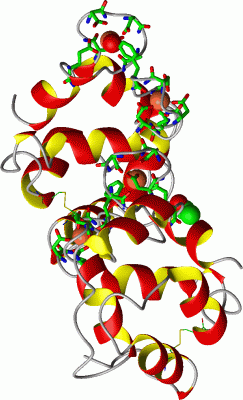
Asymmetric UnitChain A from PDB Type:PROTEIN Length:106 aligned with PRVB_MERMR | P02621 from UniProtKB/Swiss-Prot Length:108 Alignment length:106 12 22 32 42 52 62 72 82 92 102 PRVB_MERMR 3 AGILADADCAAAVKACEAADSFSYKAFFAKCGLSGKSADDIKKAFVFIDQDKSGFIEEDELKLFLQVFKAGARALTDAETKAFLKAGDSDGDGAIGVEEWVALVKA 108 SCOP domains d1a75a_ A: Parvalbumin SCOP domains CATH domains 1a75A00 A:3-108 EF-hand CATH domains Pfam domains ---------------------------------------------------------------------------------------------------------- Pfam domains SAPs(SNPs) ---------------------------------------------------------------------------------------------------------- SAPs(SNPs) PROSITE (1) -----------------------------------EF_HAND_2 PDB: A:38-73 ---EF_HAND_2 PDB: A:77-108 PROSITE (1) PROSITE (2) ------------------------------------------------EF_HAND_1 --------------------------EF_HAND_1 ------ PROSITE (2) Transcript ---------------------------------------------------------------------------------------------------------- Transcript 1a75 A 3 AGILADADCAAAVKACEAADSFSYKAFFAKCGLSGKSADDIKKAFVFIDQDKSGFIEEDELKLFLQVFKAGARALTDAETKAFLKAGDSDGDGAIGVEEWVALVKA 108 12 22 32 42 52 62 72 82 92 102 Chain B from PDB Type:PROTEIN Length:109 aligned with PRVB_MERMR | P02621 from UniProtKB/Swiss-Prot Length:108 Alignment length:109 1 | 9 19 29 39 49 59 69 79 89 99 PRVB_MERMR - -AFAGILADADCAAAVKACEAADSFSYKAFFAKCGLSGKSADDIKKAFVFIDQDKSGFIEEDELKLFLQVFKAGARALTDAETKAFLKAGDSDGDGAIGVEEWVALVKA 108 SCOP domains d1a75b_ B: Parvalbumin SCOP domains CATH domains -1a75B00 B:1-108 EF-hand CATH domains Pfam domains ------------------------------------------------------------------------------------------------------------- Pfam domains SAPs(SNPs) ------------------------------------------------------------------------------------------------------------- SAPs(SNPs) PROSITE (1) --------------------------------------EF_HAND_2 PDB: B:38-73 ---EF_HAND_2 PDB: B:77-108 PROSITE (1) PROSITE (2) ---------------------------------------------------EF_HAND_1 --------------------------EF_HAND_1 ------ PROSITE (2) Transcript ------------------------------------------------------------------------------------------------------------- Transcript 1a75 B 0 xAFAGILADADCAAAVKACEAADSFSYKAFFAKCGLSGKSADDIKKAFVFIDQDKSGFIEEDELKLFLQVFKAGARALTDAETKAFLKAGDSDGDGAIGVEEWVALVKA 108 | 9 19 29 39 49 59 69 79 89 99 0-ACE
|
||||||||||||||||||||
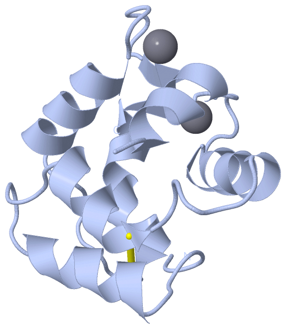
SCOP Domains (1, 2)
Asymmetric Unit
 |
CATH Domains (1, 2)
Asymmetric Unit
|
Pfam Domains (0, 0)| (no "Pfam Domain" information available for 1A75) |
Gene Ontology (2, 2)|
Asymmetric Unit(hide GO term definitions) Chain A,B (PRVB_MERMR | P02621)
|
||||||||||||||||||
Interactive Views
|
|||||||||||||||||||||||||||||||||||||||||||||||||||||||||||||||||||||||||||||||||||||||||||||||||||||||||||||||||||||||||||||||||||||||||||||||||||||||||||||||||||||||||||||||||||||||||||||||||||||
Still Images
|
||||||||||||||||
Databases
|
||||||||||||||||||||||||||||||||||||||||||||||||||||||||||||||||||||||||||||||||||||||||||||||||||||||||||||||||||||||||||||||||||||||||||||||||||||||||||||||||
Analysis Tools
|
|||||||||||||||||||||||||||||||||||||||||||||||||||||||||||||
Entries Sharing at Least One Protein Chain (UniProt ID)
Related Entries Specified in the PDB File
|
|