|
|
|
|
 Description
|
|
Compounds
|
||||||||||||||||||||||||||||||||||||||||||||||||||||
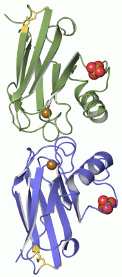
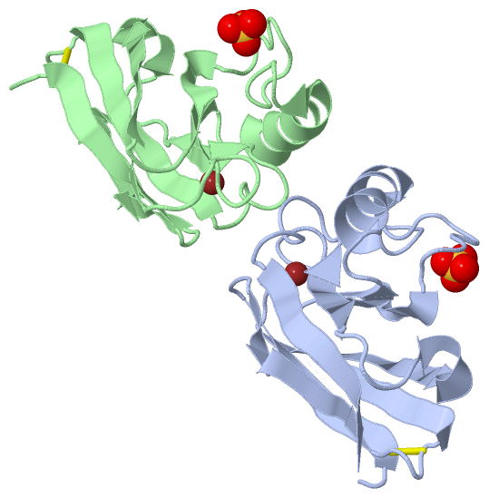
Chains, Units
Summary Information (see also Sequences/Alignments below) |
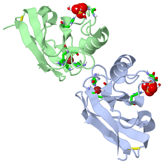
Ligands, Modified Residues, Ions (2, 4)| Asymmetric/Biological Unit (2, 4) |
Sites (4, 4)
Asymmetric Unit (4, 4)
|
SS Bonds (2, 2)
Asymmetric/Biological Unit
|
||||||||||||
Cis Peptide Bonds (0, 0)| (no "Cis Peptide Bond" information available for 1A4B) |
SAPs(SNPs)/Variants (0, 0)| (no "SAP(SNP)/Variant" information available for 1A4B) |
PROSITE Motifs (1, 2)
Asymmetric/Biological Unit (1, 2)
|
||||||||||||||||||||||||
Exons (0, 0)| (no "Exon" information available for 1A4B) |
Sequences/Alignments
Asymmetric/Biological UnitChain A from PDB Type:PROTEIN Length:129 aligned with AZUR_ACHDE | P00280 from UniProtKB/Swiss-Prot Length:149 Alignment length:129 30 40 50 60 70 80 90 100 110 120 130 140 AZUR_ACHDE 21 AQCEATIESNDAMQYNLKEMVVDKSCKQFTVHLKHVGKMAKVAMGHNWVLTKEADKQGVATDGMNAGLAQDYVKAGDTRVIAHTKVIGGGESDSVTFDVSKLTPGEAYAYFCSFPGHWAMMKGTLKLSN 149 SCOP domains d1a4ba_ A: Azurin SCOP domains CATH domains 1a4bA00 A:1-129 Cupredoxins - blue copper proteins CATH domains Pfam domains --------------------------------------------------------------------------------------------------------------------------------- Pfam domains SAPs(SNPs) --------------------------------------------------------------------------------------------------------------------------------- SAPs(SNPs) PROSITE --------------------------------------------------------------------------------------------------------COPPER_BLUE -------- PROSITE Transcript --------------------------------------------------------------------------------------------------------------------------------- Transcript 1a4b A 1 AQCEATIESNDAMQYDLKEMVVDKSCKQFTVHLKHVGKMAKSAMGHNWVLTKEADKEGVATDGMNAGLAQDYVKAGDTRVIAHTKVIGGGESDSVTFDVSKLTPGEAYAYFCSFPGHWAMHKGTLKLSN 129 10 20 30 40 50 60 70 80 90 100 110 120 Chain B from PDB Type:PROTEIN Length:129 aligned with AZUR_ACHDE | P00280 from UniProtKB/Swiss-Prot Length:149 Alignment length:129 30 40 50 60 70 80 90 100 110 120 130 140 AZUR_ACHDE 21 AQCEATIESNDAMQYNLKEMVVDKSCKQFTVHLKHVGKMAKVAMGHNWVLTKEADKQGVATDGMNAGLAQDYVKAGDTRVIAHTKVIGGGESDSVTFDVSKLTPGEAYAYFCSFPGHWAMMKGTLKLSN 149 SCOP domains d1a4bb_ B: Azurin SCOP domains CATH domains 1a4bB00 B:1-129 Cupredoxins - blue copper proteins CATH domains Pfam domains --------------------------------------------------------------------------------------------------------------------------------- Pfam domains SAPs(SNPs) --------------------------------------------------------------------------------------------------------------------------------- SAPs(SNPs) PROSITE --------------------------------------------------------------------------------------------------------COPPER_BLUE -------- PROSITE Transcript --------------------------------------------------------------------------------------------------------------------------------- Transcript 1a4b B 1 AQCEATIESNDAMQYDLKEMVVDKSCKQFTVHLKHVGKMAKSAMGHNWVLTKEADKEGVATDGMNAGLAQDYVKAGDTRVIAHTKVIGGGESDSVTFDVSKLTPGEAYAYFCSFPGHWAMHKGTLKLSN 129 10 20 30 40 50 60 70 80 90 100 110 120
|
||||||||||||||||||||
SCOP Domains (1, 2)
Asymmetric/Biological Unit
 |
CATH Domains (1, 2)
Asymmetric/Biological Unit
|
Pfam Domains (0, 0)| (no "Pfam Domain" information available for 1A4B) |
Gene Ontology (5, 5)|
Asymmetric/Biological Unit(hide GO term definitions) Chain A,B (AZUR_ACHDE | P00280)
|
||||||||||||||||||||||||||||||||||||||||||||||||
Interactive Views
|
||||||||||||||||||||||||||||||||||||||||||||||||||||||||||||||||||||||||||||||||||||||||||||||||||||||||||||||||||||||||||||||||||||||||||||||||||
Still Images
|
||||||||||||||||
Databases
|
||||||||||||||||||||||||||||||||||||||||||||||||||||||||||||||||||||||||||||||||||||||||||||||||||||||||||||||||||||||||||||||||||||||||||||||||||||||||||||||||
Analysis Tools
|
|||||||||||||||||||||||||||||||||||||||||||||||||||||||||||||
Entries Sharing at Least One Protein Chain (UniProt ID)
Related Entries Specified in the PDB File
|
|