Show PDB file:   
         Plain Text   HTML   (compressed file size)
QuickSearch:   
by PDB,NDB,UniProt,PROSITE Code or Search Term(s)  
(-)Asym./Biol. Unit - manually
(-)Asym./Biol. Unit
(-)Asym./Biol. Unit - sites
collapse expand < >
Image Asym./Biol. Unit - manually
Asym./Biol. Unit - manually  (Jmol Viewer)
Image Asym./Biol. Unit
Asym./Biol. Unit  (Jmol Viewer)
Image Asym./Biol. Unit - sites
Asym./Biol. Unit - sites  (Jmol Viewer)

(-) Description

Title :  ATP-DEPENDENT DNA LIGASE FROM BACTERIOPHAGE T7 COMPLEX WITH ATP
 
Authors :  H. S. Subramanya, A. J. Doherty, S. R. Ashford, D. B. Wigley
Date :  01 Dec 97  (Deposition) - 25 Mar 98  (Release) - 24 Feb 09  (Revision)
Method :  X-RAY DIFFRACTION
Resolution :  2.60
Chains :  Asym./Biol. Unit :  A
Keywords :  Ligase, Dna Replication (Keyword Search: [Gene Ontology, PubMed, Web (Google))
 
Reference :  H. S. Subramanya, A. J. Doherty, S. R. Ashford, D. B. Wigley
Crystal Structure Of An Atp-Dependent Dna Ligase From Bacteriophage T7.
Cell(Cambridge, Mass. ) V. 85 607 1996
PubMed-ID: 8653795  |  Reference-DOI: 10.1016/S0092-8674(00)81260-X
(for further references see the PDB file header)

(-) Compounds

Molecule 1 - DNA LIGASE
    ChainsA
    EC Number6.5.1.1
    EngineeredYES
    Expression SystemESCHERICHIA COLI
    Expression System GeneT7
    Expression System PlasmidT7
    Expression System StrainB834
    Expression System Taxid562
    Expression System VectorPET22
    Expression System Vector TypeT7
    GeneLIG
    MutationYES
    Organism ScientificENTEROBACTERIA PHAGE T7
    Organism Taxid10760

 Structural Features

(-) Chains, Units

  1
Asymmetric/Biological Unit A

Summary Information (see also Sequences/Alignments below)

(-) Ligands, Modified Residues, Ions  (1, 1)

Asymmetric/Biological Unit (1, 1)
No.NameCountTypeFull Name
1ATP1Ligand/IonADENOSINE-5'-TRIPHOSPHATE

(-) Sites  (1, 1)

Asymmetric Unit (1, 1)
No.NameEvidenceResiduesDescription
1AC1SOFTWAREGLU A:32 , ILE A:33 , LYS A:34 , TYR A:35 , ARG A:39 , ARG A:55 , GLU A:93 , TYR A:149 , ILE A:220 , LYS A:222 , LYS A:232 , TRP A:236 , LYS A:238 , HOH A:351 , HOH A:434 , HOH A:459BINDING SITE FOR RESIDUE ATP A 1

(-) SS Bonds  (0, 0)

(no "SS Bond" information available for 1A0I)

(-) Cis Peptide Bonds  (0, 0)

(no "Cis Peptide Bond" information available for 1A0I)

 Sequence-Structure Mapping

(-) SAPs(SNPs)/Variants  (0, 0)

(no "SAP(SNP)/Variant" information available for 1A0I)

(-) PROSITE Motifs  (3, 3)

Asymmetric/Biological Unit (3, 3)
 PROSITEUniProtKBPDB
No.IDACDescriptionIDLocationCountLocation
1DNA_LIGASE_A1PS00697 ATP-dependent DNA ligase AMP-binding site.DNLI_BPT732-40  1A:32-40
2DNA_LIGASE_A3PS50160 ATP-dependent DNA ligase family profile.DNLI_BPT7175-242  1A:175-242
3DNA_LIGASE_A2PS00333 ATP-dependent DNA ligase signature 2.DNLI_BPT7217-240  1A:217-240

(-) Exons   (0, 0)

(no "Exon" information available for 1A0I)

(-) Sequences/Alignments

Asymmetric/Biological Unit
   Reformat: Number of residues per line =  ('0' or empty: single-line sequence representation)
  Number of residues per labelling interval =   
  UniProt sequence: complete  aligned part    
   Show mapping: SCOP domains CATH domains Pfam domains Secondary structure (by author)
SAPs(SNPs) PROSITE motifs Exons
(details for a mapped element are shown in a popup box when the mouse pointer rests over it)
Chain A from PDB  Type:PROTEIN  Length:332
 aligned with DNLI_BPT7 | P00969 from UniProtKB/Swiss-Prot  Length:359

    Alignment length:348
                                    11        21        31        41        51        61        71        81        91       101       111       121       131       141       151       161       171       181       191       201       211       221       231       241       251       261       271       281       291       301       311       321       331       341        
            DNLI_BPT7     2 MNIKTNPFKAVSFVESAIKKALDNAGYLIAEIKYDGVRGNICVDNTANSYWLSRVSKTIPALEHLNGFDVRWKRLLNDDRCFYKDGFMLDGELMVKGVDFNTGSGLLRTKWTDTKNQEFHEELFVEPIRKKDKVPFKLHTGHLHIKLYAILPLHIVESGEDCDVMTLLMQEHVKNMLPLLQEYFPEIEWQAAESYEVYDMVELQQLYEQKRAEGHEGLIVKDPMCIYKRGKKSGWWKMKPENEADGIIQGLVWGTKGLANEGKVIGFEVLLESGRLVNATNISRALMDEFTETVKEATLSQWGFFSPYGIGDNDACTINPYDGWACQISYMEETPDGSLRHPSFVMFR 349
               SCOP domains d1a0ia2 A:2-240 ATP-dependent DNA ligase, N-terminal domain                                                                                                                                                                                    d1a0ia1 A:241-349 ATP-dependent DNA ligase                                                                    SCOP domains
               CATH domains 1a0iA01 A:2-36,A:193-240           1a0iA03 A:37-192 DNA ligase/mRNA capping enzyme                                                                                                             1a0iA01 A:2-36,A:193-240                        1a0iA02 A:241-349 Nucleic acid-binding proteins                                                               CATH domains
               Pfam domains ------------------------------------------------------------------------------------------------------------------------------------------------------------------------------------------------------------------------------------------------------------------------------------------------------------------------------------------------------------ Pfam domains
         Sec.struct. author .......eeeee..hhhhhhhhhhh..eeeee....eeeeeeee....eeee.......hhhhhh....hhhhhhhh........eeeeeeeeee.......hhhhhh............--------..............eeeeeeeeeehhhhhh.......hhhhhhhhhhhhhhhhhh....eeee..eeee..hhhhhhhhhhhhhh....eeee........eeeeeeeee...eeeeee...................eeee.....eeee.....hhhhhhhhhhhhhh.......--------...........eeeeee............eeeee. Sec.struct. author
                 SAPs(SNPs) ------------------------------------------------------------------------------------------------------------------------------------------------------------------------------------------------------------------------------------------------------------------------------------------------------------------------------------------------------------ SAPs(SNPs)
                PROSITE (1) ------------------------------DNA_LIGAS--------------------------------------------------------------------------------------------------------------------------------------DNA_LIGASE_A3  PDB: A:175-242 UniProt: 175-242                      ----------------------------------------------------------------------------------------------------------- PROSITE (1)
                PROSITE (2) -----------------------------------------------------------------------------------------------------------------------------------------------------------------------------------------------------------------------DNA_LIGASE_A2           ------------------------------------------------------------------------------------------------------------- PROSITE (2)
                 Transcript ------------------------------------------------------------------------------------------------------------------------------------------------------------------------------------------------------------------------------------------------------------------------------------------------------------------------------------------------------------ Transcript
                 1a0i A   2 VNIKTNPFKAVSFVESAIKKALDNAGYLIAEIKYDGVRGNICVDNTANSYWLSRVSKTIPALEHLNGFDVRWKRLLNDDRCFYKDGFMLDGELMVKGVDFNTGSGLLRTKWTDTKNQEFH--------RKKDKVPFKLHTGHLHIKLYAILPLHIVESGEDCDVMTLLMQEHVKNMLPLLQEYFPEIEWQAAESYEVYDMVELQQLYEQKRAEGHEGLIVKDPMCIYKRGKKSGWWKMKPENEADGIIQGLVWGTKGLANEGKVIGFEVLLESGRLVNATNISRALMDEFTETVKEATLSQWGFF--------DACTINPYDGWACQISYMEETPDGSLRHPSFVMFR 349
                                    11        21        31        41        51        61        71        81        91       101       111       121       131       141       151       161       171       181       191       201       211       221       231       241       251       261       271       281       291       301    |    -   |   321       331       341        
                                                                                                                                                 121      130                                                                                                                                                                             306      315                                  

   Legend:   → Mismatch (orange background)
  - → Gap (green background, '-', border residues have a numbering label)
    → Modified Residue (blue background, lower-case, 'x' indicates undefined single-letter code, labelled with number + name)
  x → Chemical Group (purple background, 'x', labelled with number + name, e.g. ACE or NH2)
  extra numbering lines below/above indicate numbering irregularities and modified residue names etc., number ends below/above '|'

 Classification and Annotation

(-) SCOP Domains  (2, 2)

Asymmetric/Biological Unit

(-) CATH Domains  (3, 3)

Asymmetric/Biological Unit
(-)
Class: Alpha Beta (26913)
(-)
Class: Mainly Beta (13760)

(-) Pfam Domains  (0, 0)

(no "Pfam Domain" information available for 1A0I)

(-) Gene Ontology  (12, 12)

Asymmetric/Biological Unit(hide GO term definitions)
Chain A   (DNLI_BPT7 | P00969)
molecular function
    GO:0005524    ATP binding    Interacting selectively and non-covalently with ATP, adenosine 5'-triphosphate, a universally important coenzyme and enzyme regulator.
    GO:0003677    DNA binding    Any molecular function by which a gene product interacts selectively and non-covalently with DNA (deoxyribonucleic acid).
    GO:0003910    DNA ligase (ATP) activity    Catalysis of the reaction: ATP + deoxyribonucleotide(n) + deoxyribonucleotide(m) = AMP + diphosphate + deoxyribonucleotide(n+m).
    GO:0003909    DNA ligase activity    Catalysis of the formation of a phosphodiester bond between the 3'-hydroxyl group at the end of one DNA chain and the 5'-phosphate group at the end of another. This reaction requires an energy source such as ATP or NAD+.
    GO:0016874    ligase activity    Catalysis of the joining of two substances, or two groups within a single molecule, with the concomitant hydrolysis of the diphosphate bond in ATP or a similar triphosphate.
    GO:0046872    metal ion binding    Interacting selectively and non-covalently with any metal ion.
    GO:0000166    nucleotide binding    Interacting selectively and non-covalently with a nucleotide, any compound consisting of a nucleoside that is esterified with (ortho)phosphate or an oligophosphate at any hydroxyl group on the ribose or deoxyribose.
biological process
    GO:0051103    DNA ligation involved in DNA repair    The re-formation of a broken phosphodiester bond in the DNA backbone, carried out by DNA ligase, that contributes to DNA repair.
    GO:0006310    DNA recombination    Any process in which a new genotype is formed by reassortment of genes resulting in gene combinations different from those that were present in the parents. In eukaryotes genetic recombination can occur by chromosome assortment, intrachromosomal recombination, or nonreciprocal interchromosomal recombination. Intrachromosomal recombination occurs by crossing over. In bacteria it may occur by genetic transformation, conjugation, transduction, or F-duction.
    GO:0006281    DNA repair    The process of restoring DNA after damage. Genomes are subject to damage by chemical and physical agents in the environment (e.g. UV and ionizing radiations, chemical mutagens, fungal and bacterial toxins, etc.) and by free radicals or alkylating agents endogenously generated in metabolism. DNA is also damaged because of errors during its replication. A variety of different DNA repair pathways have been reported that include direct reversal, base excision repair, nucleotide excision repair, photoreactivation, bypass, double-strand break repair pathway, and mismatch repair pathway.
    GO:0006260    DNA replication    The cellular metabolic process in which a cell duplicates one or more molecules of DNA. DNA replication begins when specific sequences, known as origins of replication, are recognized and bound by initiation proteins, and ends when the original DNA molecule has been completely duplicated and the copies topologically separated. The unit of replication usually corresponds to the genome of the cell, an organelle, or a virus. The template for replication can either be an existing DNA molecule or RNA.
    GO:0006974    cellular response to DNA damage stimulus    Any process that results in a change in state or activity of a cell (in terms of movement, secretion, enzyme production, gene expression, etc.) as a result of a stimulus indicating damage to its DNA from environmental insults or errors during metabolism.
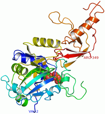
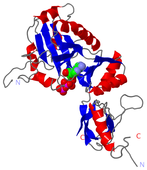
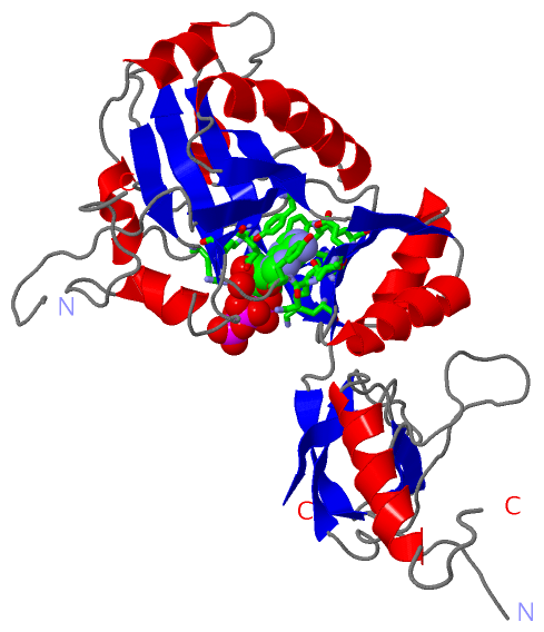
 Visualization

(-) Interactive Views

Asymmetric/Biological Unit
  Complete Structure
    Jena3D(integrated viewing of ligand, site, SAP, PROSITE, SCOP information)
    WebMol | AstexViewer[tm]@PDBe
(Java Applets, require no local installation except for Java; loading may be slow)
    STRAP
(Java WebStart application, automatic local installation, requires Java; full application with system access!)
    RasMol
(require local installation)
    Molscript (VRML)
(requires installation of a VRML viewer; select preferred view via VRML and generate a mono or stereo PDF format file)
 
  Ligands, Modified Residues, Ions
    ATP  [ RasMol | Jena3D ]  +environment [ RasMol | Jena3D ]
 
  Sites
    AC1  [ RasMol ]  +environment [ RasMol ]
 
  Cis Peptide Bonds
(no "Cis Peptide Bonds" information available for 1a0i)
 

(-) Still Images

Jmol
  protein: cartoon or spacefill or dots and stick; nucleic acid: cartoon and stick; ligands: spacefill; active site: stick
Molscript
  protein, nucleic acid: cartoon; ligands: spacefill; active site: ball and stick

 Databases and Analysis Tools

(-) Databases

Access by PDB/NDB ID
  1a0i
    Family and Domain InformationProDom | SYSTERS
    General Structural InformationGlycoscienceDB | MMDB | NDB | OCA | PDB | PDBe | PDBj | PDBsum | PDBWiki | PQS | PROTEOPEDIA
    Orientation in MembranesOPM
    Protein SurfaceSURFACE
    Secondary StructureDSSP (structure derived) | HSSP (homology derived)
    Structural GenomicsGeneCensus
    Structural NeighboursCE | VAST
    Structure ClassificationCATH | Dali | SCOP
    Validation and Original DataBMRB Data View | BMRB Restraints Grid | EDS | PROCHECK | RECOORD | WHAT_CHECK
 
Access by UniProt ID/Accession number
  DNLI_BPT7 | P00969
    Comparative Protein Structure ModelsModBase
    Genomic InformationEnsembl
    Protein-protein InteractionDIP
    Sequence, Family and Domain InformationInterPro | Pfam | SMART | UniProtKB/SwissProt
 
Access by Enzyme Classificator   (EC Number)
  6.5.1.1
    General Enzyme InformationBRENDA | EC-PDB | Enzyme | IntEnz
    PathwayKEGG | MetaCyc
 
Access by Disease Identifier   (MIM ID)
  (no 'MIM ID' available)
    Disease InformationOMIM
 
Access by GenAge ID
  (no 'GenAge ID' available)
    Age Related InformationGenAge

(-) Analysis Tools

Access by PDB/NDB ID
    Domain InformationXDom
    Interatomic Contacts of Structural UnitsCSU
    Ligand-protein ContactsLPC
    Protein CavitiescastP
    Sequence and Secondary StructurePDBCartoon
    Structure AlignmentSTRAP(Java WebStart application, automatic local installation, requires Java; full application with system access!)
    Structure and Sequence BrowserSTING
 
Access by UniProt ID/Accession number
  DNLI_BPT7 | P00969
    Protein Disorder PredictionDisEMBL | FoldIndex | GLOBPLOT (for more information see DisProt)

 Related Entries

(-) Entries Sharing at Least One Protein Chain (UniProt ID)

(no "Entries Sharing at Least One Protein Chain" available for 1A0I)

(-) Related Entries Specified in the PDB File

(no "Related Entries Specified in the PDB File" available for 1A0I)