| No. | Name | Evidence | Residues | Description |
|---|
| 01 | AC1 | SOFTWARE | DDA B:2 , DDA B:4 , MG A:13 , DG B:9 , DC B:10 | BINDING SITE FOR RESIDUE CRH B 3 |
| 02 | AC2 | SOFTWARE | DDA A:8 , DDA A:10 , MG A:13 , DG A:3 , DC A:4 | BINDING SITE FOR RESIDUE CRH A 9 |
| 03 | AC3 | SOFTWARE | DDA B:2 , DG B:9 | BINDING SITE FOR RESIDUE DDA B 1 |
| 04 | AC4 | SOFTWARE | DDA B:1 , CRH B:3 , DDL A:11 | BINDING SITE FOR RESIDUE DDA B 2 |
| 05 | AC5 | SOFTWARE | CRH B:3 , DDL B:5 , DG B:11 , DA B:12 | BINDING SITE FOR RESIDUE DDA B 4 |
| 06 | AC6 | SOFTWARE | DDA A:7 , CRH A:9 | BINDING SITE FOR RESIDUE DDA A 8 |
| 07 | AC7 | SOFTWARE | CRH A:9 , DDL A:11 , DG A:5 , DA A:6 | BINDING SITE FOR RESIDUE DDA A 10 |
| 08 | AC8 | SOFTWARE | DDA A:8 , DG A:3 | BINDING SITE FOR RESIDUE DDA A 7 |
| 09 | AC9 | SOFTWARE | DDA B:4 , MDA B:6 , DA B:12 | BINDING SITE FOR RESIDUE DDL B 5 |
| 10 | AD1 | SOFTWARE | DDA B:2 , DDA A:10 , MDA A:12 , DA A:6 | BINDING SITE FOR RESIDUE DDL A 11 |
| 11 | AD2 | SOFTWARE | DDL B:5 , DG B:11 , DA B:12 | BINDING SITE FOR RESIDUE MDA B 6 |
| 12 | AD3 | SOFTWARE | DDL A:11 , DG A:5 , DA A:6 | BINDING SITE FOR RESIDUE MDA A 12 |
| 13 | AD4 | SOFTWARE | CRH B:3 , CRH A:9 | BINDING SITE FOR RESIDUE MG A 13 |
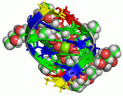
 Description
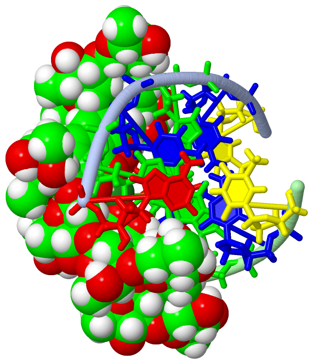
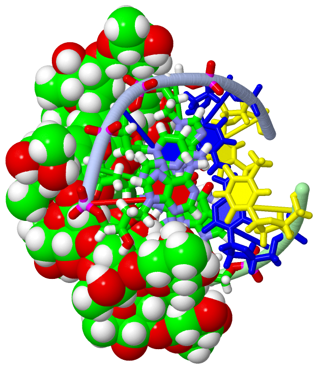
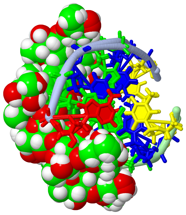