Show PDB file:   
         Plain Text   HTML   (compressed file size)
QuickSearch:   
by PDB,NDB,UniProt,PROSITE Code or Search Term(s)  
(-)Asym./Biol. Unit
(-)Asym./Biol. Unit - sites
collapse expand < >
Image Asym./Biol. Unit
Asym./Biol. Unit  (Jmol Viewer)
Image Asym./Biol. Unit - sites
Asym./Biol. Unit - sites  (Jmol Viewer)

(-) Description

Title :  COMPLEX STRUCTURE OF JMJD5 AND SUBSTRATE
 
Authors :  H. L. Liu, Y. Wang, C. Wang, G. Y. Zhang
Date :  14 Dec 15  (Deposition) - 14 Dec 16  (Release) - 14 Dec 16  (Revision)
Method :  X-RAY DIFFRACTION
Resolution :  2.42
Chains :  Asym./Biol. Unit :  A
Keywords :  Histone Enzyme, Oxidoreductase (Keyword Search: [Gene Ontology, PubMed, Web (Google))
 
Reference :  H. L. Liu, Y. Wang, C. Wang, S. D. Dai, G. Y. Zhang
To Be Published
To Be Published
PubMed: search

(-) Compounds

Molecule 1 - LYSINE-SPECIFIC DEMETHYLASE 8
    ChainsA
    EC Number1.14.11.27
    EngineeredYES
    Expression SystemESCHERICHIA COLI
    Expression System PlasmidPGEX4T1
    Expression System StrainBL21(DE3)
    Expression System Taxid469008
    Expression System Vector TypePLASMID
    GeneKDM8, JMJD5
    Organism CommonHUMAN
    Organism ScientificHOMO SAPIENS
    Organism Taxid9606
    SynonymJMJC DOMAIN-CONTAINING PROTEIN 5,JUMONJI DOMAIN-CONTAINING PROTEIN 5

 Structural Features

(-) Chains, Units

  1
Asymmetric/Biological Unit A

Summary Information (see also Sequences/Alignments below)
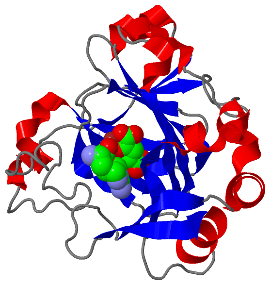
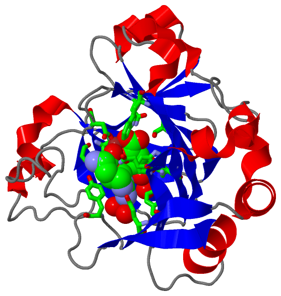
(-) Ligands, Modified Residues, Ions  (3, 3)

Asymmetric/Biological Unit (3, 3)
No.NameCountTypeFull Name
1AKG1Ligand/Ion2-OXOGLUTARIC ACID
2NMM1Ligand/Ion(2S)-2-AMINO-5-[(N-METHYLCARBAMIMIDOYL)AMINO]PENTANOICACID
3ZN1Ligand/IonZINC ION

(-) Sites  (3, 3)

Asymmetric Unit (3, 3)
No.NameEvidenceResiduesDescription
1AC1SOFTWARETYR A:272 , TRP A:310 , SER A:318 , HIS A:321 , ASP A:323 , ASN A:327 , LYS A:336 , HIS A:400 , VAL A:402 , SER A:412 , TRP A:414 , NMM A:502 , ZN A:503 , HOH A:648binding site for residue AKG A 501
2AC2SOFTWAREGLU A:238 , TYR A:243 , TYR A:272 , GLN A:275 , TRP A:310 , SER A:318 , HIS A:321 , ASP A:323 , TRP A:414 , AKG A:501 , ZN A:503 , HOH A:632 , HOH A:633binding site for residue NMM A 502
3AC3SOFTWAREHIS A:321 , ASP A:323 , HIS A:400 , AKG A:501 , NMM A:502binding site for residue ZN A 503

(-) SS Bonds  (0, 0)

(no "SS Bond" information available for 5FBJ)

(-) Cis Peptide Bonds  (0, 0)

(no "Cis Peptide Bond" information available for 5FBJ)

 Sequence-Structure Mapping

(-) SAPs(SNPs)/Variants  (0, 0)

(no "SAP(SNP)/Variant" information available for 5FBJ)

(-) PROSITE Motifs  (0, 0)

(no "PROSITE Motif" information available for 5FBJ)

(-) Exons   (0, 0)

(no "Exon" information available for 5FBJ)

(-) Sequences/Alignments

Asymmetric/Biological Unit
   Reformat: Number of residues per line =  ('0' or empty: single-line sequence representation)
  Number of residues per labelling interval =   
  UniProt sequence: complete  aligned part    
   Show mapping: SCOP domains CATH domains Pfam domains Secondary structure (by author)
SAPs(SNPs) PROSITE motifs Exons
(details for a mapped element are shown in a popup box when the mouse pointer rests over it)
Chain A from PDB  Type:PROTEIN  Length:236
                                                                                                                                                                                                                                                                            
               SCOP domains -------------------------------------------------------------------------------------------------------------------------------------------------------------------------------------------------------------------------------------------- SCOP domains
               CATH domains -------------------------------------------------------------------------------------------------------------------------------------------------------------------------------------------------------------------------------------------- CATH domains
               Pfam domains -------------------------------------------------------------------------------------------------------------------------------------------------------------------------------------------------------------------------------------------- Pfam domains
         Sec.struct. author .ee..eee..hhhhhhhhh......eeee......hhhhhhhhhhhhhhhh..eeeeee.........eeeeeehhhhhhhhh.......eeeeeehhhhhhhhhhh....hhhhhhh..hhhhheeeeeee....eeeee....eeeeeeee..eeeeeehhhhhhhhh......................hhhhh....eeeee....eeee....eeeeee...eeeeeeee. Sec.struct. author
                 SAPs(SNPs) -------------------------------------------------------------------------------------------------------------------------------------------------------------------------------------------------------------------------------------------- SAPs(SNPs)
                    PROSITE -------------------------------------------------------------------------------------------------------------------------------------------------------------------------------------------------------------------------------------------- PROSITE
                 Transcript -------------------------------------------------------------------------------------------------------------------------------------------------------------------------------------------------------------------------------------------- Transcript
                 5fbj A 181 GSTVPRLHRPSLQHFREQFLVPGRPVILKGVADHWPCMQKWSLEYIQEIAGCRTVPVEVGSRYTDEEWSQTLMTVNEFISKYIVNEPRDVGYLAQHQLFDQIPELKQDISIPDYCSLGDGEEEEITINAWFGPQGTISPLHQDPQQNFLVQVMGRKYIRLYSPQESGALYPHDTHLLHNTSQVDVENPDLEKFPKFAKAPFLSCILSPGEILFIPVKYWHYVRALDLSFSVSFWWS 416
                                   190       200       210       220       230       240       250       260       270       280       290       300       310       320       330       340       350       360       370       380       390       400       410      

   Legend:   → Mismatch (orange background)
  - → Gap (green background, '-', border residues have a numbering label)
    → Modified Residue (blue background, lower-case, 'x' indicates undefined single-letter code, labelled with number + name)
  x → Chemical Group (purple background, 'x', labelled with number + name, e.g. ACE or NH2)
  extra numbering lines below/above indicate numbering irregularities and modified residue names etc., number ends below/above '|'

 Classification and Annotation

(-) SCOP Domains  (0, 0)

(no "SCOP Domain" information available for 5FBJ)

(-) CATH Domains  (0, 0)

(no "CATH Domain" information available for 5FBJ)

(-) Pfam Domains  (0, 0)

(no "Pfam Domain" information available for 5FBJ)

(-) Gene Ontology  (13, 13)

Asymmetric/Biological Unit(hide GO term definitions)

 Visualization

(-) Interactive Views

Asymmetric/Biological Unit
  Complete Structure
    Jena3D(integrated viewing of ligand, site, SAP, PROSITE, SCOP information)
    WebMol | AstexViewer[tm]@PDBe
(Java Applets, require no local installation except for Java; loading may be slow)
    STRAP
(Java WebStart application, automatic local installation, requires Java; full application with system access!)
    RasMol
(require local installation)
    Molscript (VRML)
(requires installation of a VRML viewer; select preferred view via VRML and generate a mono or stereo PDF format file)
 
  Ligands, Modified Residues, Ions
    AKG  [ RasMol | Jena3D ]  +environment [ RasMol | Jena3D ]
    NMM  [ RasMol | Jena3D ]  +environment [ RasMol | Jena3D ]
    ZN  [ RasMol | Jena3D ]  +environment [ RasMol | Jena3D ]
 
  Sites
    AC1  [ RasMol ]  +environment [ RasMol ]
    AC2  [ RasMol ]  +environment [ RasMol ]
    AC3  [ RasMol ]  +environment [ RasMol ]
 
  Cis Peptide Bonds
(no "Cis Peptide Bonds" information available for 5fbj)
 

(-) Still Images

Jmol
  protein: cartoon or spacefill or dots and stick; nucleic acid: cartoon and stick; ligands: spacefill; active site: stick
Molscript
  protein, nucleic acid: cartoon; ligands: spacefill; active site: ball and stick

 Databases and Analysis Tools

(-) Databases

Access by PDB/NDB ID
  5fbj
    Family and Domain InformationProDom | SYSTERS
    General Structural InformationGlycoscienceDB | MMDB | NDB | OCA | PDB | PDBe | PDBj | PDBsum | PDBWiki | PQS | PROTEOPEDIA
    Orientation in MembranesOPM
    Protein SurfaceSURFACE
    Secondary StructureDSSP (structure derived) | HSSP (homology derived)
    Structural GenomicsGeneCensus
    Structural NeighboursCE | VAST
    Structure ClassificationCATH | Dali | SCOP
    Validation and Original DataBMRB Data View | BMRB Restraints Grid | EDS | PROCHECK | RECOORD | WHAT_CHECK
 
Access by UniProt ID/Accession number
  KDM8_HUMAN | Q8N371
    Comparative Protein Structure ModelsModBase
    Genomic InformationEnsembl
    Protein-protein InteractionDIP
    Sequence, Family and Domain InformationInterPro | Pfam | SMART | UniProtKB/SwissProt
 
Access by Enzyme Classificator   (EC Number)
  1.14.11.27
    General Enzyme InformationBRENDA | EC-PDB | Enzyme | IntEnz
    PathwayKEGG | MetaCyc
 
Access by Disease Identifier   (MIM ID)
  (no 'MIM ID' available)
    Disease InformationOMIM
 
Access by GenAge ID
  (no 'GenAge ID' available)
    Age Related InformationGenAge

(-) Analysis Tools

Access by PDB/NDB ID
    Domain InformationXDom
    Interatomic Contacts of Structural UnitsCSU
    Ligand-protein ContactsLPC
    Protein CavitiescastP
    Sequence and Secondary StructurePDBCartoon
    Structure AlignmentSTRAP(Java WebStart application, automatic local installation, requires Java; full application with system access!)
    Structure and Sequence BrowserSTING
 
Access by UniProt ID/Accession number
  KDM8_HUMAN | Q8N371
    Protein Disorder PredictionDisEMBL | FoldIndex | GLOBPLOT (for more information see DisProt)

 Related Entries

(-) Entries Sharing at Least One Protein Chain (UniProt ID)

UniProtKB/Swiss-Prot
        KDM8_HUMAN | Q8N3713uyj 4aap 4gaz 4gjy 4gjz 4qu1

(-) Related Entries Specified in the PDB File

4qu1 CRYSTAL STRUCTURE OF HUMAN JMJD5 JMJ-C DOMAIN