|
|
|
|
 Description
|
|
Compounds
|
||||||||||||||||||||||||||||||||||||||||||||||||||||||||||||
Chains, Units
Summary Information (see also Sequences/Alignments below) |
Ligands, Modified Residues, Ions (4, 7)| Asymmetric/Biological Unit (4, 7) |
Sites (7, 7)
Asymmetric Unit (7, 7)
|
SS Bonds (0, 0)| (no "SS Bond" information available for 5EUU) |
Cis Peptide Bonds (0, 0)| (no "Cis Peptide Bond" information available for 5EUU) |
SAPs(SNPs)/Variants (0, 0)| (no "SAP(SNP)/Variant" information available for 5EUU) |
PROSITE Motifs (0, 0)| (no "PROSITE Motif" information available for 5EUU) |
Exons (0, 0)| (no "Exon" information available for 5EUU) |
Sequences/Alignments
Asymmetric/Biological Unit
Chain A from PDB Type:PROTEIN Length:129
SCOP domains --------------------------------------------------------------------------------------------------------------------------------- SCOP domains
CATH domains --------------------------------------------------------------------------------------------------------------------------------- CATH domains
Pfam domains --------------------------------------------------------------------------------------------------------------------------------- Pfam domains
SAPs(SNPs) --------------------------------------------------------------------------------------------------------------------------------- SAPs(SNPs)
PROSITE --------------------------------------------------------------------------------------------------------------------------------- PROSITE
Transcript --------------------------------------------------------------------------------------------------------------------------------- Transcript
5euu A 505 SPSYTVLGQLPDTDVYIDIDAYEEVKEIPGIKIFQINAPIYYANSDLYNIHTVILDFTQVNFMDSVGVKTLAGIVKEYGDVGIYVYLAGCSAQVVNDLTSNRFFENPALKELLFHSIHDAVLGSQVREA 718
514 524 534 544 639 649 659 669 679 689 699 709
552|
638
|
||||||||||||||||||||
SCOP Domains (0, 0)| (no "SCOP Domain" information available for 5EUU) |
CATH Domains (0, 0)| (no "CATH Domain" information available for 5EUU) |
Pfam Domains (0, 0)| (no "Pfam Domain" information available for 5EUU) |
Gene Ontology (42, 42)|
Asymmetric/Biological Unit(hide GO term definitions) |
Interactive Views
|
|||||||||||||||||||||||||||||||||||||||||||||||||||||||||||||||||||||||||||||||||||||||||||||||||||||||||||||||||||||||||||||||||||||||||||||||||||||||||||||||||||||||||||||||||||||
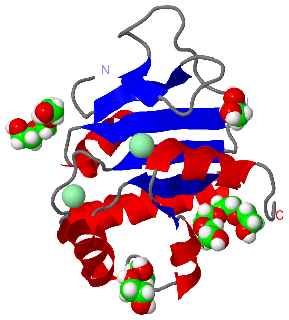
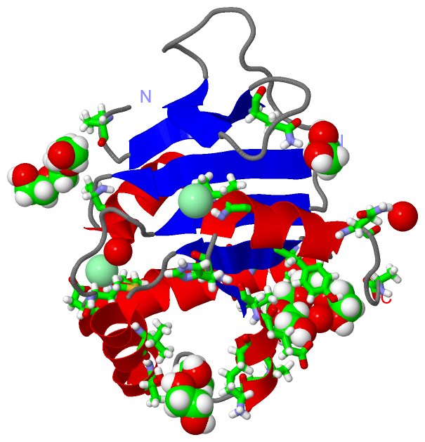
Still Images
|
||||||||||||||||
Databases
|
||||||||||||||||||||||||||||||||||||||||||||||||||||||||||||||||||||||||||||||||||||||||||||||||||||||||||||||||||||||||||||||||||||||||||||||||||||||||||||||||
Analysis Tools
|
|||||||||||||||||||||||||||||||||||||||||||||||||||||||||||||
Entries Sharing at Least One Protein Chain (UniProt ID)
Related Entries Specified in the PDB File
|
|