Show PDB file:   
         Plain Text   HTML   (compressed file size)
QuickSearch:   
by PDB,NDB,UniProt,PROSITE Code or Search Term(s)  
(-)Asym./Biol. Unit
(-)Asym./Biol. Unit - sites
collapse expand < >
Image Asym./Biol. Unit
Asym./Biol. Unit  (Jmol Viewer)
Image Asym./Biol. Unit - sites
Asym./Biol. Unit - sites  (Jmol Viewer)

(-) Description

Title :  X-RAY CRYSTAL STRUCTURE OF THE DIMERIC BMP ANTAGONIST NBL1
 
Authors :  T. B. Thompson, K. Nolan, C. Kattamuri
Date :  24 Nov 14  (Deposition) - 14 Jan 15  (Release) - 17 Aug 16  (Revision)
Method :  X-RAY DIFFRACTION
Resolution :  2.50
Chains :  Asym./Biol. Unit :  A,B
Keywords :  Bmp Antagonist, Dan Family, Cystine-Knot, Bmp Binding Protein (Keyword Search: [Gene Ontology, PubMed, Web (Google))
 
Reference :  K. Nolan, C. Kattamuri, D. M. Luedeke, E. B. Angerman, S. A. Rankin, M. L. Stevens, A. M. Zorn, T. B. Thompson
Structure Of Neuroblastoma Suppressor Of Tumorigenicity 1 (Nbl1): Insights For The Functional Variability Across Bone Morphogenetic Protein (Bmp) Antagonists.
J. Biol. Chem. V. 290 4759 2015
PubMed-ID: 25561725  |  Reference-DOI: 10.1074/JBC.M114.628412

(-) Compounds

Molecule 1 - NEUROBLASTOMA SUPPRESSOR OF TUMORIGENICITY 1
    ChainsA, B
    EngineeredYES
    Expression SystemESCHERICHIA COLI
    Expression System Taxid562
    FragmentUNP RESIDUES 52-167
    GeneNBL1, DAN, DAND1
    Organism CommonHUMAN
    Organism ScientificHOMO SAPIENS
    Organism Taxid9606
    SynonymDAN DOMAIN FAMILY MEMBER 1,PROTEIN N03,ZINC FINGER PROTEIN DAN

 Structural Features

(-) Chains, Units

  12
Asymmetric/Biological Unit AB

Summary Information (see also Sequences/Alignments below)

(-) Ligands, Modified Residues, Ions  (1, 2)

Asymmetric/Biological Unit (1, 2)
No.NameCountTypeFull Name
1NAG2Ligand/IonN-ACETYL-D-GLUCOSAMINE

(-) Sites  (2, 2)

Asymmetric Unit (2, 2)
No.NameEvidenceResiduesDescription
1AC1SOFTWAREASN A:39 , ARG A:57binding site for Mono-Saccharide NAG A 501 bound to ASN A 39
2AC2SOFTWAREASN B:39binding site for Mono-Saccharide NAG B 501 bound to ASN B 39

(-) SS Bonds  (10, 10)

Asymmetric/Biological Unit
No.Residues
1A:35 -A:85
2A:49 -A:99
3A:59 -A:118
4A:63 -A:120
5A:82 -A:123
6B:35 -B:85
7B:49 -B:99
8B:59 -B:118
9B:63 -B:120
10B:82 -B:123

(-) Cis Peptide Bonds  (2, 2)

Asymmetric/Biological Unit
No.Residues
1Gly A:45 -His A:46
2Cys A:99 -Pro A:100

 Sequence-Structure Mapping

(-) SAPs(SNPs)/Variants  (0, 0)

(no "SAP(SNP)/Variant" information available for 4X1J)

(-) PROSITE Motifs  (0, 0)

(no "PROSITE Motif" information available for 4X1J)

(-) Exons   (0, 0)

(no "Exon" information available for 4X1J)

(-) Sequences/Alignments

Asymmetric/Biological Unit
   Reformat: Number of residues per line =  ('0' or empty: single-line sequence representation)
  Number of residues per labelling interval =   
  UniProt sequence: complete  aligned part    
   Show mapping: SCOP domains CATH domains Pfam domains Secondary structure (by author)
SAPs(SNPs) PROSITE motifs Exons
(details for a mapped element are shown in a popup box when the mouse pointer rests over it)
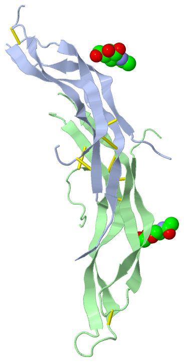
Chain A from PDB  Type:PROTEIN  Length:83
                                                                                                                   
               SCOP domains ----------------------------------------------------------------------------------- SCOP domains
               CATH domains ----------------------------------------------------------------------------------- CATH domains
               Pfam domains ----------------------------------------------------------------------------------- Pfam domains
         Sec.struct. author ....eeeeeeeeee.........eeeeeeeeeeeeeee...ee.eeeeeeeeeeeeeeee......eeeeeeeeeeeeee... Sec.struct. author
                 SAPs(SNPs) ----------------------------------------------------------------------------------- SAPs(SNPs)
                    PROSITE ----------------------------------------------------------------------------------- PROSITE
                 Transcript ----------------------------------------------------------------------------------- Transcript
                 4x1j A  30 DKSAWCEAKNITQIVGHSGCEAKSIQNRACLGQCFSYSVPNTFVHCDSCMPAQSMWEIVTLECPGVDKLVEKILHCSCQACGK 125
                                    39        49        59        69  ||    86        96    || 112       122   
                                                                     72|                  101|                 
                                                                      80                   108                 

Chain B from PDB  Type:PROTEIN  Length:94
                                                                                                                              
               SCOP domains ---------------------------------------------------------------------------------------------- SCOP domains
               CATH domains ---------------------------------------------------------------------------------------------- CATH domains
               Pfam domains ---------------------------------------------------------------------------------------------- Pfam domains
         Sec.struct. author ....eeeeeeeeeee.......eeeeeeeeeeeeeeee.....eeeeeeeeeeeeeeeeeee........eeeeeeeeeeeeeeee........ Sec.struct. author
                 SAPs(SNPs) ---------------------------------------------------------------------------------------------- SAPs(SNPs)
                    PROSITE ---------------------------------------------------------------------------------------------- PROSITE
                 Transcript ---------------------------------------------------------------------------------------------- Transcript
                 4x1j B  30 DKSAWCEAKNITQIVGHSGCEAKSIQNRACLGQCFSYSVPESLVHCDSCMPAQSMWEIVTLECPGHEEVPRVDKLVEKILHCSCQACGKEPSHE 130
                                    39        49        59        69|       86        96       106       116       126    
                                                                  69|                                                     
                                                                   77                                                     

   Legend:   → Mismatch (orange background)
  - → Gap (green background, '-', border residues have a numbering label)
    → Modified Residue (blue background, lower-case, 'x' indicates undefined single-letter code, labelled with number + name)
  x → Chemical Group (purple background, 'x', labelled with number + name, e.g. ACE or NH2)
  extra numbering lines below/above indicate numbering irregularities and modified residue names etc., number ends below/above '|'

 Classification and Annotation

(-) SCOP Domains  (0, 0)

(no "SCOP Domain" information available for 4X1J)

(-) CATH Domains  (0, 0)

(no "CATH Domain" information available for 4X1J)

(-) Pfam Domains  (0, 0)

(no "Pfam Domain" information available for 4X1J)

(-) Gene Ontology  (14, 14)

Asymmetric/Biological Unit(hide GO term definitions)

 Visualization

(-) Interactive Views

Asymmetric/Biological Unit
  Complete Structure
    Jena3D(integrated viewing of ligand, site, SAP, PROSITE, SCOP information)
    WebMol | AstexViewer[tm]@PDBe
(Java Applets, require no local installation except for Java; loading may be slow)
    STRAP
(Java WebStart application, automatic local installation, requires Java; full application with system access!)
    RasMol
(require local installation)
    Molscript (VRML)
(requires installation of a VRML viewer; select preferred view via VRML and generate a mono or stereo PDF format file)
 
  Ligands, Modified Residues, Ions
    NAG  [ RasMol | Jena3D ]  +environment [ RasMol | Jena3D ]
 
  Sites
    AC1  [ RasMol ]  +environment [ RasMol ]
    AC2  [ RasMol ]  +environment [ RasMol ]
 
  Cis Peptide Bonds
    Cys A:99 - Pro A:100   [ RasMol ]  
    Gly A:45 - His A:46   [ RasMol ]  
 

(-) Still Images

Jmol
  protein: cartoon or spacefill or dots and stick; nucleic acid: cartoon and stick; ligands: spacefill; active site: stick
Molscript
  protein, nucleic acid: cartoon; ligands: spacefill; active site: ball and stick

 Databases and Analysis Tools

(-) Databases

Access by PDB/NDB ID
  4x1j
    Family and Domain InformationProDom | SYSTERS
    General Structural InformationGlycoscienceDB | MMDB | NDB | OCA | PDB | PDBe | PDBj | PDBsum | PDBWiki | PQS | PROTEOPEDIA
    Orientation in MembranesOPM
    Protein SurfaceSURFACE
    Secondary StructureDSSP (structure derived) | HSSP (homology derived)
    Structural GenomicsGeneCensus
    Structural NeighboursCE | VAST
    Structure ClassificationCATH | Dali | SCOP
    Validation and Original DataBMRB Data View | BMRB Restraints Grid | EDS | PROCHECK | RECOORD | WHAT_CHECK
 
Access by UniProt ID/Accession number
  NBL1_HUMAN | P41271
    Comparative Protein Structure ModelsModBase
    Genomic InformationEnsembl
    Protein-protein InteractionDIP
    Sequence, Family and Domain InformationInterPro | Pfam | SMART | UniProtKB/SwissProt
 
Access by Enzyme Classificator   (EC Number)
  (no 'Enzyme Classificator' available)
    General Enzyme InformationBRENDA | EC-PDB | Enzyme | IntEnz
    PathwayKEGG | MetaCyc
 
Access by Disease Identifier   (MIM ID)
  (no 'MIM ID' available)
    Disease InformationOMIM
 
Access by GenAge ID
  (no 'GenAge ID' available)
    Age Related InformationGenAge

(-) Analysis Tools

Access by PDB/NDB ID
    Domain InformationXDom
    Interatomic Contacts of Structural UnitsCSU
    Ligand-protein ContactsLPC
    Protein CavitiescastP
    Sequence and Secondary StructurePDBCartoon
    Structure AlignmentSTRAP(Java WebStart application, automatic local installation, requires Java; full application with system access!)
    Structure and Sequence BrowserSTING
 
Access by UniProt ID/Accession number
  NBL1_HUMAN | P41271
    Protein Disorder PredictionDisEMBL | FoldIndex | GLOBPLOT (for more information see DisProt)

 Related Entries

(-) Entries Sharing at Least One Protein Chain (UniProt ID)

UniProtKB/Swiss-Prot
        NBL1_HUMAN | P412714yu8

(-) Related Entries Specified in the PDB File

4jph THE RELATED ENTRY IS A PROTEIN THAT IS A PART OF THE SAME PROTEIN FAMILY.