Show PDB file:   
         Plain Text   HTML   (compressed file size)
QuickSearch:   
by PDB,NDB,UniProt,PROSITE Code or Search Term(s)  
(-)Asymmetric Unit
(-)Asym. Unit - sites
(-)Biological Unit 1
(-)Biol. Unit 1 - sites
collapse expand < >
Image Asymmetric Unit
Asymmetric Unit  (Jmol Viewer)
Image Asym. Unit - sites
Asym. Unit - sites  (Jmol Viewer)
Image Biological Unit 1
Biological Unit 1  (Jmol Viewer)
Image Biol. Unit 1 - sites
Biol. Unit 1 - sites  (Jmol Viewer)

(-) Description

Title :  CRYSTAL STRUCTURE OF TRYPANOSOMA BRUCEI DUTPASE WITH DUMP, PLANAR [ALF3-OPO3] TRANSITION STATE ANALOGUE, MG2+, AND NA+
 
Authors :  G. R. Hemsworth, D. Gonzalez-Pacanowska, K. S. Wilson
Date :  06 Feb 12  (Deposition) - 07 Aug 13  (Release) - 06 Nov 13  (Revision)
Method :  X-RAY DIFFRACTION
Resolution :  1.70
Chains :  Asym. Unit :  A
Biol. Unit 1:  A  (2x)
Keywords :  All Alpha Ntp Pyrophosphohydrolase, All Alpha Ntp Pyrophosphatase, Hydrolase (Keyword Search: [Gene Ontology, PubMed, Web (Google))
 
Reference :  G. R. Hemsworth, D. Gonzalez-Pacanowska, K. S. Wilson
On The Catalytic Mechanism Of Dimeric Dutpases.
Biochem. J. V. 456 81 2013
PubMed-ID: 24001052  |  Reference-DOI: 10.1042/BJ20130796

(-) Compounds

Molecule 1 - DEOXYURIDINE TRIPHOSPHATASE
    ChainsA
    EC Number3.6.1.23
    EngineeredYES
    Expression SystemESCHERICHIA COLI
    Expression System PlasmidPET-YSBLLIC
    Expression System StrainBL21(DE3)
    Expression System Taxid469008
    Expression System Vector TypePLASMID
    GeneTB927.7.5160
    Organism ScientificTRYPANOSOMA BRUCEI
    Organism Taxid5691
    Strain427
    SynonymDEOXYURIDINE TRIPHOSPHATE NUCLEOTIDOHYDROLASE

 Structural Features

(-) Chains, Units

  1
Asymmetric Unit A
Biological Unit 1 (2x)A

Summary Information (see also Sequences/Alignments below)
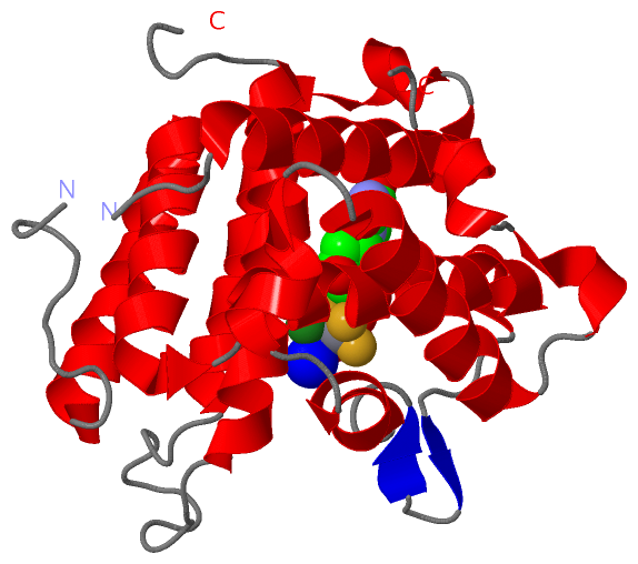
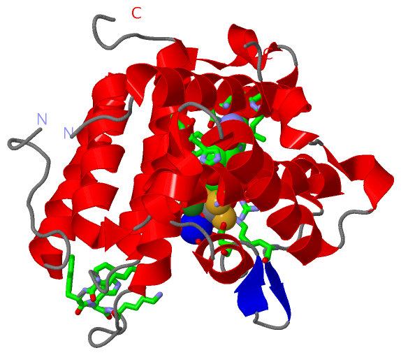
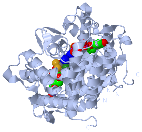
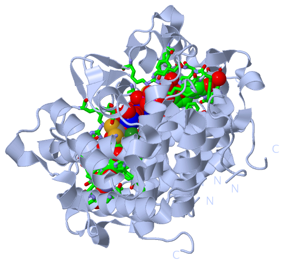
(-) Ligands, Modified Residues, Ions  (5, 6)

Asymmetric Unit (5, 6)
No.NameCountTypeFull Name
1AF31Ligand/IonALUMINUM FLUORIDE
2MG2Ligand/IonMAGNESIUM ION
3NA1Ligand/IonSODIUM ION
4PO41Ligand/IonPHOSPHATE ION
5UMP1Ligand/Ion2'-DEOXYURIDINE 5'-MONOPHOSPHATE
Biological Unit 1 (3, 6)
No.NameCountTypeFull Name
1AF32Ligand/IonALUMINUM FLUORIDE
2MG-1Ligand/IonMAGNESIUM ION
3NA-1Ligand/IonSODIUM ION
4PO42Ligand/IonPHOSPHATE ION
5UMP2Ligand/Ion2'-DEOXYURIDINE 5'-MONOPHOSPHATE

(-) Sites  (6, 6)

Asymmetric Unit (6, 6)
No.NameEvidenceResiduesDescription
1AC1SOFTWAREGLN A:21 , LEU A:24 , ASN A:25 , GLU A:48 , LYS A:59 , TRP A:60 , TRP A:61 , ASP A:79 , HIS A:82 , PHE A:83 , ASN A:212 , ARG A:215 , TYR A:220 , LYS A:227 , AF3 A:302 , PO4 A:303 , MG A:304 , HOH A:404 , HOH A:405 , HOH A:437BINDING SITE FOR RESIDUE UMP A 301
2AC2SOFTWAREGLU A:48 , GLU A:51 , ASP A:79 , LYS A:208 , ARG A:215 , LYS A:227 , ASN A:235 , UMP A:301 , PO4 A:303 , MG A:304 , MG A:305 , NA A:306 , HOH A:401 , HOH A:485BINDING SITE FOR RESIDUE AF3 A 302
3AC3SOFTWAREGLU A:51 , LYS A:59 , LYS A:62 , LYS A:227 , UMP A:301 , AF3 A:302 , MG A:304 , NA A:306 , HOH A:401 , HOH A:437 , HOH A:485BINDING SITE FOR RESIDUE PO4 A 303
4AC4SOFTWAREGLU A:48 , GLU A:51 , UMP A:301 , AF3 A:302 , PO4 A:303 , MG A:305 , HOH A:437BINDING SITE FOR RESIDUE MG A 304
5AC5SOFTWAREGLU A:48 , GLU A:51 , GLU A:76 , ASP A:79 , AF3 A:302 , MG A:304 , NA A:306 , HOH A:401BINDING SITE FOR RESIDUE MG A 305
6AC6SOFTWAREGLU A:51 , GLU A:76 , AF3 A:302 , PO4 A:303 , MG A:305 , HOH A:401 , HOH A:402 , HOH A:403 , HOH A:485BINDING SITE FOR RESIDUE NA A 306

(-) SS Bonds  (0, 0)

(no "SS Bond" information available for 4DL8)

(-) Cis Peptide Bonds  (1, 1)

Asymmetric Unit
No.Residues
1Phe A:159 -Pro A:160

 Sequence-Structure Mapping

(-) SAPs(SNPs)/Variants  (0, 0)

(no "SAP(SNP)/Variant" information available for 4DL8)

(-) PROSITE Motifs  (0, 0)

(no "PROSITE Motif" information available for 4DL8)

(-) Exons   (0, 0)

(no "Exon" information available for 4DL8)

(-) Sequences/Alignments

Asymmetric Unit
   Reformat: Number of residues per line =  ('0' or empty: single-line sequence representation)
  Number of residues per labelling interval =   
  UniProt sequence: complete  aligned part    
   Show mapping: SCOP domains CATH domains Pfam domains Secondary structure (by author)
SAPs(SNPs) PROSITE motifs Exons
(details for a mapped element are shown in a popup box when the mouse pointer rests over it)
Chain A from PDB  Type:PROTEIN  Length:224
 aligned with Q57ZH3_TRYB2 | Q57ZH3 from UniProtKB/TrEMBL  Length:287

    Alignment length:281
                                    16        26        36        46        56        66        76        86        96       106       116       126       136       146       156       166       176       186       196       206       216       226       236       246       256       266       276       286 
         Q57ZH3_TRYB2     7 VSLSPLILRSLAELQDGLNTVVDKNWRQLRRPGDWSLAITMEAAELLDSYPWKWWKNVKAQPDLQNVKIELTDILHFSLSGAMQVSDENSGAVHKAEAGSNGESGKHWCYFDQPRALPAAGGAEYVACVETPGSSLSAPVSADECDLADFMFFPLSDTNNALASFQNIIRLASLQRFQLVTSAVIAAADDIGFNLVAYYVAKHTLNGIRQMKGYKDGTYVKVQKGVEDNELLHGCISPFSLDDVTNEGNYKTKWDDIMHRVYDAFGTPKEERLNIGHWLKS 287
               SCOP domains ----------------------------------------------------------------------------------------------------------------------------------------------------------------------------------------------------------------------------------------------------------------------------------------- SCOP domains
               CATH domains ----------------------------------------------------------------------------------------------------------------------------------------------------------------------------------------------------------------------------------------------------------------------------------------- CATH domains
               Pfam domains ----------------------------------------------------------------------------------------------------------------------------------------------------------------------------------------------------------------------------------------------------------------------------------------- Pfam domains
         Sec.struct. author ...hhhhhhhhhhhhhhhhhhhh.hhhhhhhhhhhhhhhhhhhhhhhh..............hhhhhhhhhhhhhhhhhhhhh......---------------------------------------------------------..........hhhhhhhhhhhhhhhhhh.hhhhhhhhhhhhhhhhh.hhhhhhhhhhhhhhhhhhhhhhhh....ee..ee...hhhhhhh..hhhhhhh..hhhhhhhhhhhhhhhhh..hhhhh.hhhhh... Sec.struct. author
                 SAPs(SNPs) ----------------------------------------------------------------------------------------------------------------------------------------------------------------------------------------------------------------------------------------------------------------------------------------- SAPs(SNPs)
                    PROSITE ----------------------------------------------------------------------------------------------------------------------------------------------------------------------------------------------------------------------------------------------------------------------------------------- PROSITE
                 Transcript ----------------------------------------------------------------------------------------------------------------------------------------------------------------------------------------------------------------------------------------------------------------------------------------- Transcript
                 4dl8 A   7 VSLSPLILRSLAELQDGLNTVVDKNWRQLRRPGDWSLAITMEAAELLDSYPWKWWKNVKAQPDLQNVKIELTDILHFSLSGAMQVSDEN---------------------------------------------------------LADFMFFPLSDTNNALASFQNIIRLASLQRFQLVTSAVIAAADDIGFNLVAYYVAKHTLNGIRQMKGYKDGTYVKVQKGVEDNELLHGCISPFSLDDVTNEGNYKTKWDDIMHRVYDAFGTPKEERLNIGHWLKS 287
                                    16        26        36        46        56        66        76        86        |-         -         -         -         -         -      |156       166       176       186       196       206       216       226       236       246       256       266       276       286 
                                                                                                                   95                                                       153                                                                                                                                      

   Legend:   → Mismatch (orange background)
  - → Gap (green background, '-', border residues have a numbering label)
    → Modified Residue (blue background, lower-case, 'x' indicates undefined single-letter code, labelled with number + name)
  x → Chemical Group (purple background, 'x', labelled with number + name, e.g. ACE or NH2)
  extra numbering lines below/above indicate numbering irregularities and modified residue names etc., number ends below/above '|'

 Classification and Annotation

(-) SCOP Domains  (0, 0)

(no "SCOP Domain" information available for 4DL8)

(-) CATH Domains  (0, 0)

(no "CATH Domain" information available for 4DL8)

(-) Pfam Domains  (0, 0)

(no "Pfam Domain" information available for 4DL8)

(-) Gene Ontology  (3, 3)

Asymmetric Unit(hide GO term definitions)
Chain A   (Q57ZH3_TRYB2 | Q57ZH3)
molecular function
    GO:0004170    dUTP diphosphatase activity    Catalysis of the reaction: dUTP + H2O = dUMP + diphosphate.
    GO:0016787    hydrolase activity    Catalysis of the hydrolysis of various bonds, e.g. C-O, C-N, C-C, phosphoric anhydride bonds, etc. Hydrolase is the systematic name for any enzyme of EC class 3.
    GO:0046872    metal ion binding    Interacting selectively and non-covalently with any metal ion.

 Visualization

(-) Interactive Views

Asymmetric Unit
  Complete Structure
    Jena3D(integrated viewing of ligand, site, SAP, PROSITE, SCOP information)
    WebMol | AstexViewer[tm]@PDBe
(Java Applets, require no local installation except for Java; loading may be slow)
    STRAP
(Java WebStart application, automatic local installation, requires Java; full application with system access!)
    RasMol
(require local installation)
    Molscript (VRML)
(requires installation of a VRML viewer; select preferred view via VRML and generate a mono or stereo PDF format file)
 
  Ligands, Modified Residues, Ions
    AF3  [ RasMol | Jena3D ]  +environment [ RasMol | Jena3D ]
    MG  [ RasMol | Jena3D ]  +environment [ RasMol | Jena3D ]
    NA  [ RasMol | Jena3D ]  +environment [ RasMol | Jena3D ]
    PO4  [ RasMol | Jena3D ]  +environment [ RasMol | Jena3D ]
    UMP  [ RasMol | Jena3D ]  +environment [ RasMol | Jena3D ]
 
  Sites
    AC1  [ RasMol ]  +environment [ RasMol ]
    AC2  [ RasMol ]  +environment [ RasMol ]
    AC3  [ RasMol ]  +environment [ RasMol ]
    AC4  [ RasMol ]  +environment [ RasMol ]
    AC5  [ RasMol ]  +environment [ RasMol ]
    AC6  [ RasMol ]  +environment [ RasMol ]
 
  Cis Peptide Bonds
    Phe A:159 - Pro A:160   [ RasMol ]  
 
Biological Unit
  Complete Structure
    Biological Unit 1  [ Jena3D ]

(-) Still Images

Jmol
  protein: cartoon or spacefill or dots and stick; nucleic acid: cartoon and stick; ligands: spacefill; active site: stick
Molscript
  protein, nucleic acid: cartoon; ligands: spacefill; active site: ball and stick

 Databases and Analysis Tools

(-) Databases

Access by PDB/NDB ID
  4dl8
    Family and Domain InformationProDom | SYSTERS
    General Structural InformationGlycoscienceDB | MMDB | NDB | OCA | PDB | PDBe | PDBj | PDBsum | PDBWiki | PQS | PROTEOPEDIA
    Orientation in MembranesOPM
    Protein SurfaceSURFACE
    Secondary StructureDSSP (structure derived) | HSSP (homology derived)
    Structural GenomicsGeneCensus
    Structural NeighboursCE | VAST
    Structure ClassificationCATH | Dali | SCOP
    Validation and Original DataBMRB Data View | BMRB Restraints Grid | EDS | PROCHECK | RECOORD | WHAT_CHECK
 
Access by UniProt ID/Accession number
  Q57ZH3_TRYB2 | Q57ZH3
    Comparative Protein Structure ModelsModBase
    Genomic InformationEnsembl
    Protein-protein InteractionDIP
    Sequence, Family and Domain InformationInterPro | Pfam | SMART | UniProtKB/TrEMBL
 
Access by Enzyme Classificator   (EC Number)
  3.6.1.23
    General Enzyme InformationBRENDA | EC-PDB | Enzyme | IntEnz
    PathwayKEGG | MetaCyc
 
Access by Disease Identifier   (MIM ID)
  (no 'MIM ID' available)
    Disease InformationOMIM
 
Access by GenAge ID
  (no 'GenAge ID' available)
    Age Related InformationGenAge

(-) Analysis Tools

Access by PDB/NDB ID
    Domain InformationXDom
    Interatomic Contacts of Structural UnitsCSU
    Ligand-protein ContactsLPC
    Protein CavitiescastP
    Sequence and Secondary StructurePDBCartoon
    Structure AlignmentSTRAP(Java WebStart application, automatic local installation, requires Java; full application with system access!)
    Structure and Sequence BrowserSTING
 
Access by UniProt ID/Accession number
  Q57ZH3_TRYB2 | Q57ZH3
    Protein Disorder PredictionDisEMBL | FoldIndex | GLOBPLOT (for more information see DisProt)

 Related Entries

(-) Entries Sharing at Least One Protein Chain (UniProt ID)

UniProtKB/TrEMBL
        Q57ZH3_TRYB2 | Q57ZH34dk2 4dk4 4dkb 4dlc

(-) Related Entries Specified in the PDB File

4dk2 4dk4 4dkb 4dlc