| molecular function |
|---|
| | GO:0005509 | | calcium ion binding | | Interacting selectively and non-covalently with calcium ions (Ca2+). |
| | GO:0047485 | | protein N-terminus binding | | Interacting selectively and non-covalently with a protein N-terminus, the end of any peptide chain at which the 2-amino (or 2-imino) function of a constituent amino acid is not attached in peptide linkage to another amino-acid residue. |
| | GO:0005515 | | protein binding | | Interacting selectively and non-covalently with any protein or protein complex (a complex of two or more proteins that may include other nonprotein molecules). |
| | GO:0032403 | | protein complex binding | | Interacting selectively and non-covalently with any protein complex (a complex of two or more proteins that may include other nonprotein molecules). |
| biological process |
|---|
| | GO:0051017 | | actin filament bundle assembly | | The assembly of actin filament bundles; actin filaments are on the same axis but may be oriented with the same or opposite polarities and may be packed with different levels of tightness. |
| | GO:0007015 | | actin filament organization | | A process that is carried out at the cellular level which results in the assembly, arrangement of constituent parts, or disassembly of cytoskeletal structures comprising actin filaments. Includes processes that control the spatial distribution of actin filaments, such as organizing filaments into meshworks, bundles, or other structures, as by cross-linking. |
| | GO:0007628 | | adult walking behavior | | The behavior of an adult relating to the progression of that organism along the ground by the process of lifting and setting down each leg. |
| | GO:0042491 | | auditory receptor cell differentiation | | The process in which a relatively unspecialized cell acquires specialized features of an auditory hair cell. |
| | GO:0060088 | | auditory receptor cell stereocilium organization | | A process that is carried out at the cellular level which results in the assembly, arrangement of constituent parts, or disassembly of a stereocilium. A stereocilium is an actin-based protrusion from the apical surface of auditory hair cells. |
| | GO:0007155 | | cell adhesion | | The attachment of a cell, either to another cell or to an underlying substrate such as the extracellular matrix, via cell adhesion molecules. |
| | GO:0050973 | | detection of mechanical stimulus involved in equilibrioception | | The series of events involved in equilibrioception in which a mechanical stimulus is received and converted into a molecular signal. During equilibrioception, mechanical stimuli may be in the form of input from pressure receptors or from the labyrinth system of the inner ears. |
| | GO:0050910 | | detection of mechanical stimulus involved in sensory perception of sound | | The series of events involved in the perception of sound vibration in which the vibration is received and converted into a molecular signal. |
| | GO:0050957 | | equilibrioception | | The series of events required for an organism to receive an orientational stimulus, convert it to a molecular signal, and recognize and characterize the signal. Equilibrioception refers to a combination of processes by which an organism can perceive its orientation with respect to gravity. In animals, stimuli come from labyrinth system of the inner ears, monitoring the direction of motion; visual stimuli, with information on orientation and motion; pressure receptors, which tell the organism which body surfaces are in contact with the ground; and proprioceptive cues, which report which parts of the body are in motion. |
| | GO:0007156 | | homophilic cell adhesion via plasma membrane adhesion molecules | | The attachment of a plasma membrane adhesion molecule in one cell to an identical molecule in an adjacent cell. |
| | GO:0048839 | | inner ear development | | The process whose specific outcome is the progression of the inner ear over time, from its formation to the mature structure. |
| | GO:0060122 | | inner ear receptor stereocilium organization | | A process that is carried out at the cellular level which results in the assembly, arrangement of constituent parts, or disassembly of a stereocilium. A stereocilium is an actin-based protrusion from the apical surface of inner ear receptor cells. |
| | GO:0007626 | | locomotory behavior | | The specific movement from place to place of an organism in response to external or internal stimuli. Locomotion of a whole organism in a manner dependent upon some combination of that organism's internal state and external conditions. |
| | GO:0002009 | | morphogenesis of an epithelium | | The process in which the anatomical structures of epithelia are generated and organized. An epithelium consists of closely packed cells arranged in one or more layers, that covers the outer surfaces of the body or lines any internal cavity or tube. |
| | GO:0035264 | | multicellular organism growth | | The increase in size or mass of an entire multicellular organism, as opposed to cell growth. |
| | GO:0045494 | | photoreceptor cell maintenance | | Any process preventing the degeneration of the photoreceptor, a specialized cell type that is sensitive to light. |
| | GO:0051592 | | response to calcium ion | | Any process that results in a change in state or activity of a cell or an organism (in terms of movement, secretion, enzyme production, gene expression, etc.) as a result of a calcium ion stimulus. |
| | GO:0060013 | | righting reflex | | A reflex process in which an animal immediately tries to turn over after being placed in a supine position. |
| | GO:0050953 | | sensory perception of light stimulus | | The series of events required for an organism to receive a sensory light stimulus, convert it to a molecular signal, and recognize and characterize the signal. This is a neurological process. |
| | GO:0007605 | | sensory perception of sound | | The series of events required for an organism to receive an auditory stimulus, convert it to a molecular signal, and recognize and characterize the signal. Sonic stimuli are detected in the form of vibrations and are processed to form a sound. |
| | GO:0001964 | | startle response | | An action or movement due to the application of a sudden unexpected stimulus. |
| | GO:0007601 | | visual perception | | The series of events required for an organism to receive a visual stimulus, convert it to a molecular signal, and recognize and characterize the signal. Visual stimuli are detected in the form of photons and are processed to form an image. |
| cellular component |
|---|
| | GO:0005737 | | cytoplasm | | All of the contents of a cell excluding the plasma membrane and nucleus, but including other subcellular structures. |
| | GO:0070062 | | extracellular exosome | | A vesicle that is released into the extracellular region by fusion of the limiting endosomal membrane of a multivesicular body with the plasma membrane. Extracellular exosomes, also simply called exosomes, have a diameter of about 40-100 nm. |
| | GO:0005576 | | extracellular region | | The space external to the outermost structure of a cell. For cells without external protective or external encapsulating structures this refers to space outside of the plasma membrane. This term covers the host cell environment outside an intracellular parasite. |
| | GO:0005615 | | extracellular space | | That part of a multicellular organism outside the cells proper, usually taken to be outside the plasma membranes, and occupied by fluid. |
| | GO:0016021 | | integral component of membrane | | The component of a membrane consisting of the gene products and protein complexes having at least some part of their peptide sequence embedded in the hydrophobic region of the membrane. |
| | GO:0016020 | | membrane | | A lipid bilayer along with all the proteins and protein complexes embedded in it an attached to it. |
| | GO:0001750 | | photoreceptor outer segment | | The outer segment of a vertebrate photoreceptor that contains discs of photoreceptive membranes. |
| | GO:0005886 | | plasma membrane | | The membrane surrounding a cell that separates the cell from its external environment. It consists of a phospholipid bilayer and associated proteins. |
| | GO:0032420 | | stereocilium | | An actin-based protrusion from the apical surface of auditory and vestibular hair cells and of neuromast cells. These protrusions are supported by a bundle of cross-linked actin filaments (an actin cable), oriented such that the plus (barbed) ends are at the tip of the protrusion, capped by a tip complex which bridges to the plasma. Bundles of stereocilia act as mechanosensory organelles. |
| | GO:0032421 | | stereocilium bundle | | A bundle of cross-linked stereocilia, arranged around a kinocilium on the apical surface of a sensory hair cell (e.g. a neuromast, auditory or vestibular hair cell). Stereocilium bundles act as mechanosensory organelles by responding to fluid motion or fluid pressure changes. |
| | GO:0045202 | | synapse | | The junction between a nerve fiber of one neuron and another neuron, muscle fiber or glial cell. As the nerve fiber approaches the synapse it enlarges into a specialized structure, the presynaptic nerve ending, which contains mitochondria and synaptic vesicles. At the tip of the nerve ending is the presynaptic membrane; facing it, and separated from it by a minute cleft (the synaptic cleft) is a specialized area of membrane on the receiving cell, known as the postsynaptic membrane. In response to the arrival of nerve impulses, the presynaptic nerve ending secretes molecules of neurotransmitters into the synaptic cleft. These diffuse across the cleft and transmit the signal to the postsynaptic membrane. |
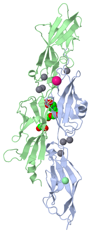
 Description
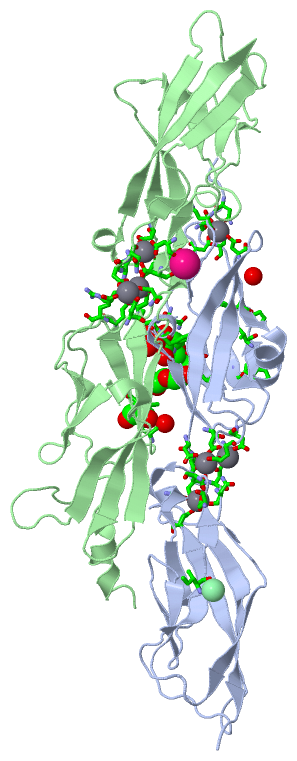