| molecular function |
|---|
| | GO:0016787 | | hydrolase activity | | Catalysis of the hydrolysis of various bonds, e.g. C-O, C-N, C-C, phosphoric anhydride bonds, etc. Hydrolase is the systematic name for any enzyme of EC class 3. |
| | GO:0016740 | | transferase activity | | Catalysis of the transfer of a group, e.g. a methyl group, glycosyl group, acyl group, phosphorus-containing, or other groups, from one compound (generally regarded as the donor) to another compound (generally regarded as the acceptor). Transferase is the systematic name for any enzyme of EC class 2. |
| | GO:0016757 | | transferase activity, transferring glycosyl groups | | Catalysis of the transfer of a glycosyl group from one compound (donor) to another (acceptor). |
| biological process |
|---|
| | GO:0071555 | | cell wall organization | | A process that results in the assembly, arrangement of constituent parts, or disassembly of the cell wall, the rigid or semi-rigid envelope lying outside the cell membrane of plant, fungal and most prokaryotic cells, maintaining their shape and protecting them from osmotic lysis. |
| | GO:0009252 | | peptidoglycan biosynthetic process | | The chemical reactions and pathways resulting in the formation of peptidoglycans, any of a class of glycoconjugates found in bacterial cell walls. |
| | GO:0008360 | | regulation of cell shape | | Any process that modulates the surface configuration of a cell. |
| | GO:0030435 | | sporulation resulting in formation of a cellular spore | | The process in which a relatively unspecialized cell acquires the specialized features of a cellular spore, a cell form that can be used for dissemination, for survival of adverse conditions because of its heat and dessication resistance, and/or for reproduction. |
| cellular component |
|---|
| | GO:0031160 | | spore wall | | The specialized envelope lying outside the cell membrane of a spore. |
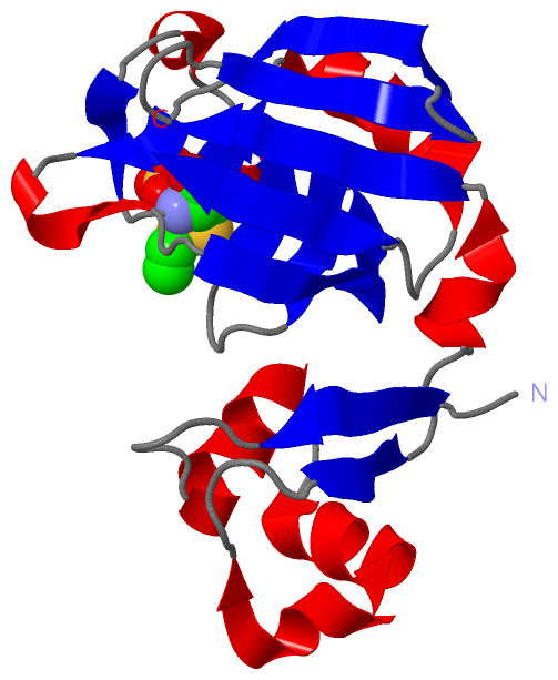
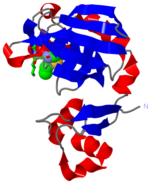
 Description
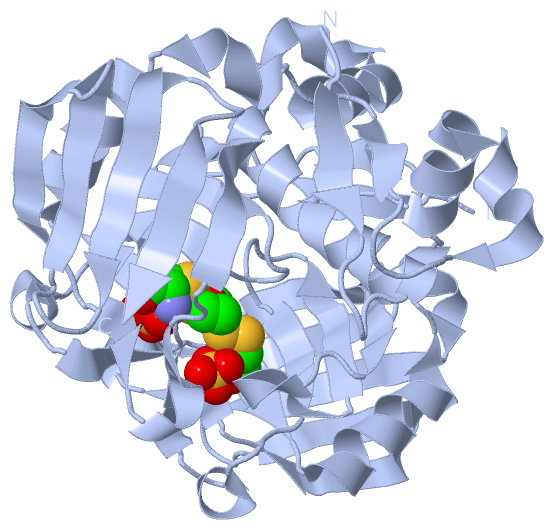
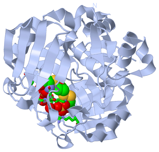