Show PDB file:   
         Plain Text   HTML   (compressed file size)
QuickSearch:   
by PDB,NDB,UniProt,PROSITE Code or Search Term(s)  
(-)Asymmetric Unit
(-)Biological Unit 1
(-)Biological Unit 2
(-)Biological Unit 3
collapse expand < >
Image Asymmetric Unit
Asymmetric Unit  (Jmol Viewer)
Image Biological Unit 1
Biological Unit 1  (Jmol Viewer)
Image Biological Unit 2
Biological Unit 2  (Jmol Viewer)
Image Biological Unit 3
Biological Unit 3  (Jmol Viewer)

(-) Description

Title :  CRYSTAL STRUCTURE OF PAR3-NTD DOMAIN
 
Authors :  W. Wang, F. Gao, W. Gong, F. Sun, W. Feng
Date :  29 Nov 12  (Deposition) - 17 Jul 13  (Release) - 17 Jul 13  (Revision)
Method :  X-RAY DIFFRACTION
Resolution :  2.90
Chains :  Asym. Unit :  A,B
Biol. Unit 1:  A,B  (1x)
Biol. Unit 2:  A  (1x)
Biol. Unit 3:  B  (1x)
Keywords :  Pb1 Like Motif, Duf3534, Cell Polarity Protein, Signaling Protein (Keyword Search: [Gene Ontology, PubMed, Web (Google))
 
Reference :  Y. Zhang, W. Wang, J. Chen, K. Zhang, F. Gao, B. Gao, S. Zhang, M. Dong, F. Besenbacher, W. Gong, M. Zhang, F. Sun, W. Feng
Structural Insights Into The Intrinsic Self-Assembly Of Par-3 N-Terminal Domain.
Structure V. 21 997 2013
PubMed-ID: 23643951  |  Reference-DOI: 10.1016/J.STR.2013.04.004

(-) Compounds

Molecule 1 - PARTITIONING DEFECTIVE 3 HOMOLOG
    ChainsA, B
    EngineeredYES
    Expression SystemESCHERICHIA COLI
    Expression System PlasmidPET32A
    Expression System StrainBL21-CODON PLUS
    Expression System Taxid511693
    Expression System Vector TypePLASMID
    FragmentN-TERMINAL DOMAIN OF PAR3
    GenePARD3, PAR3
    Organism CommonBROWN RAT,RAT,RATS
    Organism ScientificRATTUS NORVEGICUS
    Organism Taxid10116
    SynonymPAR-3, PARD-3, ATYPICAL PKC ISOTYPE-SPECIFIC-INTERACTING PROTEIN, ASIP, ATYPICAL PKC-SPECIFIC-BINDING PROTEIN, ASBP

 Structural Features

(-) Chains, Units

  12
Asymmetric Unit AB
Biological Unit 1 (1x)AB
Biological Unit 2 (1x)A 
Biological Unit 3 (1x) B

Summary Information (see also Sequences/Alignments below)

(-) Ligands, Modified Residues, Ions  (0, 0)

(no "Ligand,Modified Residues,Ions" information available for 4I6P)

(-) Sites  (0, 0)

(no "Site" information available for 4I6P)

(-) SS Bonds  (0, 0)

(no "SS Bond" information available for 4I6P)

(-) Cis Peptide Bonds  (1, 1)
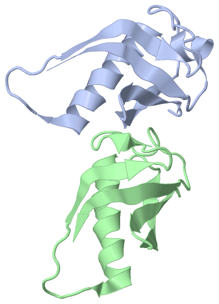
Asymmetric Unit
No.Residues
1Ala A:39 -Lys A:40

 Sequence-Structure Mapping

(-) SAPs(SNPs)/Variants  (0, 0)

(no "SAP(SNP)/Variant" information available for 4I6P)

(-) PROSITE Motifs  (0, 0)

(no "PROSITE Motif" information available for 4I6P)

(-) Exons   (0, 0)

(no "Exon" information available for 4I6P)

(-) Sequences/Alignments

Asymmetric Unit
   Reformat: Number of residues per line =  ('0' or empty: single-line sequence representation)
  Number of residues per labelling interval =   
  UniProt sequence: complete  aligned part    
   Show mapping: SCOP domains CATH domains Pfam domains Secondary structure (by author)
SAPs(SNPs) PROSITE motifs Exons
(details for a mapped element are shown in a popup box when the mouse pointer rests over it)
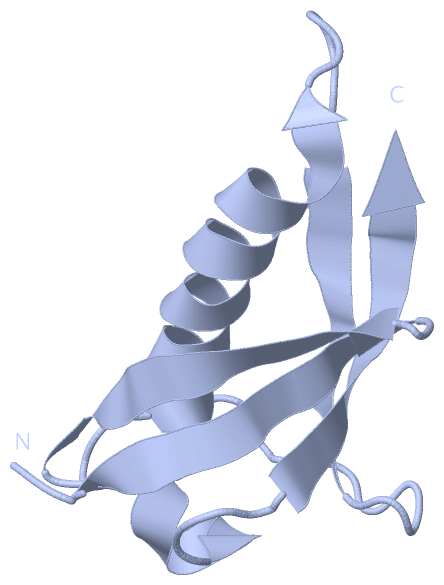
Chain A from PDB  Type:PROTEIN  Length:84
                                                                                                                   
               SCOP domains ------------------------------------------------------------------------------------ SCOP domains
               CATH domains ------------------------------------------------------------------------------------ CATH domains
               Pfam domains ------------------------------------------------------------------------------------ Pfam domains
         Sec.struct. author ..eeeeeee..eeeeeeee....hhhhhhhhhhhhhhhhhh......eeeeeee...........hhhhhh....eeeeeeee. Sec.struct. author
                 SAPs(SNPs) ------------------------------------------------------------------------------------ SAPs(SNPs)
                    PROSITE ------------------------------------------------------------------------------------ PROSITE
                 Transcript ------------------------------------------------------------------------------------ Transcript
                  4i6p A -1 SEFKVTVCFGRTRVVVPCGDGRMKVFSLIQQAVTRYRKAVAKDPNYWIQVHRLEHGDGGILDLDDILCDVADDKDRLVAVFDEQ 82
                                     8        18        28        38        48        58        68        78    

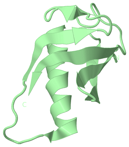
Chain B from PDB  Type:PROTEIN  Length:82
                                                                                                                 
               SCOP domains ---------------------------------------------------------------------------------- SCOP domains
               CATH domains ---------------------------------------------------------------------------------- CATH domains
               Pfam domains ---------------------------------------------------------------------------------- Pfam domains
         Sec.struct. author .eeeeeee..eeeeeeee....hhhhhhhhhhhhhhhhhh.......eeeeee....eee..............eeeeeee. Sec.struct. author
                 SAPs(SNPs) ---------------------------------------------------------------------------------- SAPs(SNPs)
                    PROSITE ---------------------------------------------------------------------------------- PROSITE
                 Transcript ---------------------------------------------------------------------------------- Transcript
                  4i6p B  0 EFKVTVCFGRTRVVVPCGDGRMKVFSLIQQAVTRYRKAVAKDPNYWIQVHRLEHGDGGILDLDDILCDVADDKDRLVAVFDE 81
                                     9        19        29        39        49        59        69        79  

   Legend:   → Mismatch (orange background)
  - → Gap (green background, '-', border residues have a numbering label)
    → Modified Residue (blue background, lower-case, 'x' indicates undefined single-letter code, labelled with number + name)
  x → Chemical Group (purple background, 'x', labelled with number + name, e.g. ACE or NH2)
  extra numbering lines below/above indicate numbering irregularities and modified residue names etc., number ends below/above '|'

 Classification and Annotation

(-) SCOP Domains  (0, 0)

(no "SCOP Domain" information available for 4I6P)

(-) CATH Domains  (0, 0)

(no "CATH Domain" information available for 4I6P)

(-) Pfam Domains  (0, 0)

(no "Pfam Domain" information available for 4I6P)

(-) Gene Ontology  (30, 30)

Asymmetric Unit(hide GO term definitions)

 Visualization

(-) Interactive Views

Asymmetric Unit
  Complete Structure
    Jena3D(integrated viewing of ligand, site, SAP, PROSITE, SCOP information)
    WebMol | AstexViewer[tm]@PDBe
(Java Applets, require no local installation except for Java; loading may be slow)
    STRAP
(Java WebStart application, automatic local installation, requires Java; full application with system access!)
    RasMol
(require local installation)
    Molscript (VRML)
(requires installation of a VRML viewer; select preferred view via VRML and generate a mono or stereo PDF format file)
 
  Ligands, Modified Residues, Ions
(no "Ligands, Modified Residues, Ions" information available for 4i6p)
 
  Sites
(no "Sites" information available for 4i6p)
 
  Cis Peptide Bonds
    Ala A:39 - Lys A:40   [ RasMol ]  
 
Biological Units
  Complete Structure
    Biological Unit 1  [ Jena3D ]
    Biological Unit 2  [ Jena3D ]
    Biological Unit 3  [ Jena3D ]

(-) Still Images

Jmol
  protein: cartoon or spacefill or dots and stick; nucleic acid: cartoon and stick; ligands: spacefill; active site: stick
Molscript
  protein, nucleic acid: cartoon; ligands: spacefill; active site: ball and stick

 Databases and Analysis Tools

(-) Databases

Access by PDB/NDB ID
  4i6p
    Family and Domain InformationProDom | SYSTERS
    General Structural InformationGlycoscienceDB | MMDB | NDB | OCA | PDB | PDBe | PDBj | PDBsum | PDBWiki | PQS | PROTEOPEDIA
    Orientation in MembranesOPM
    Protein SurfaceSURFACE
    Secondary StructureDSSP (structure derived) | HSSP (homology derived)
    Structural GenomicsGeneCensus
    Structural NeighboursCE | VAST
    Structure ClassificationCATH | Dali | SCOP
    Validation and Original DataBMRB Data View | BMRB Restraints Grid | EDS | PROCHECK | RECOORD | WHAT_CHECK
 
Access by UniProt ID/Accession number
  PARD3_RAT | Q9Z340
    Comparative Protein Structure ModelsModBase
    Genomic InformationEnsembl
    Protein-protein InteractionDIP
    Sequence, Family and Domain InformationInterPro | Pfam | SMART | UniProtKB/SwissProt
 
Access by Enzyme Classificator   (EC Number)
  (no 'Enzyme Classificator' available)
    General Enzyme InformationBRENDA | EC-PDB | Enzyme | IntEnz
    PathwayKEGG | MetaCyc
 
Access by Disease Identifier   (MIM ID)
  (no 'MIM ID' available)
    Disease InformationOMIM
 
Access by GenAge ID
  (no 'GenAge ID' available)
    Age Related InformationGenAge

(-) Analysis Tools

Access by PDB/NDB ID
    Domain InformationXDom
    Interatomic Contacts of Structural UnitsCSU
    Ligand-protein ContactsLPC
    Protein CavitiescastP
    Sequence and Secondary StructurePDBCartoon
    Structure AlignmentSTRAP(Java WebStart application, automatic local installation, requires Java; full application with system access!)
    Structure and Sequence BrowserSTING
 
Access by UniProt ID/Accession number
  PARD3_RAT | Q9Z340
    Protein Disorder PredictionDisEMBL | FoldIndex | GLOBPLOT (for more information see DisProt)

 Related Entries

(-) Entries Sharing at Least One Protein Chain (UniProt ID)

UniProtKB/Swiss-Prot
        PARD3_RAT | Q9Z3402k1z 2k20 2ns5 2ogp 3zee 4dc2

(-) Related Entries Specified in the PDB File

3zee STRUCTURE OF HELICAL ASSEMBLY