| molecular function |
|---|
| | GO:0005524 | | ATP binding | | Interacting selectively and non-covalently with ATP, adenosine 5'-triphosphate, a universally important coenzyme and enzyme regulator. |
| | GO:0016887 | | ATPase activity | | Catalysis of the reaction: ATP + H2O = ADP + phosphate + 2 H+. May or may not be coupled to another reaction. |
| | GO:0042626 | | ATPase activity, coupled to transmembrane movement of substances | | Catalysis of the reaction: ATP + H2O = ADP + phosphate, to directly drive the active transport of a substance across a membrane. |
| | GO:0016787 | | hydrolase activity | | Catalysis of the hydrolysis of various bonds, e.g. C-O, C-N, C-C, phosphoric anhydride bonds, etc. Hydrolase is the systematic name for any enzyme of EC class 3. |
| | GO:0000166 | | nucleotide binding | | Interacting selectively and non-covalently with a nucleotide, any compound consisting of a nucleoside that is esterified with (ortho)phosphate or an oligophosphate at any hydroxyl group on the ribose or deoxyribose. |
| | GO:0008233 | | peptidase activity | | Catalysis of the hydrolysis of a peptide bond. A peptide bond is a covalent bond formed when the carbon atom from the carboxyl group of one amino acid shares electrons with the nitrogen atom from the amino group of a second amino acid. |
| | GO:0008565 | | protein transporter activity | | Enables the directed movement of proteins into, out of or within a cell, or between cells. |
| biological process |
|---|
| | GO:0030253 | | protein secretion by the type I secretion system | | The process in which proteins are secreted into the extracellular milieu via the type I secretion system; secretion occurs in a continuous process without the distinct presence of periplasmic intermediates and does not involve proteolytic processing of secreted proteins. |
| | GO:0006508 | | proteolysis | | The hydrolysis of proteins into smaller polypeptides and/or amino acids by cleavage of their peptide bonds. |
| | GO:0055085 | | transmembrane transport | | The process in which a solute is transported across a lipid bilayer, from one side of a membrane to the other |
| | GO:0006810 | | transport | | The directed movement of substances (such as macromolecules, small molecules, ions) or cellular components (such as complexes and organelles) into, out of or within a cell, or between cells, or within a multicellular organism by means of some agent such as a transporter, pore or motor protein. |
| cellular component |
|---|
| | GO:0016021 | | integral component of membrane | | The component of a membrane consisting of the gene products and protein complexes having at least some part of their peptide sequence embedded in the hydrophobic region of the membrane. |
| | GO:0016020 | | membrane | | A lipid bilayer along with all the proteins and protein complexes embedded in it an attached to it. |
| | GO:0005886 | | plasma membrane | | The membrane surrounding a cell that separates the cell from its external environment. It consists of a phospholipid bilayer and associated proteins. |
| | GO:0030256 | | type I protein secretion system complex | | A complex of three secretory proteins that carry out secretion in the type I secretion system: an inner membrane transport ATPase (termed ABC protein for ATP-binding cassette), which provides the energy for protein secretion; an outer membrane protein, which is exported via the sec pathway; and a membrane fusion protein, which is anchored in the inner membrane and spans the periplasmic space. |
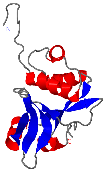
 Description
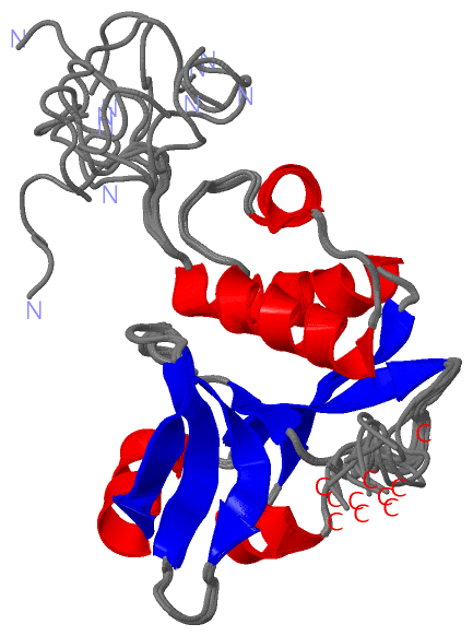