|
|
|
|
 Description
|
|
Compounds
|
||||||||||||||||||||||||||||||||||||||||||||||||||||||||
Chains, Units
Summary Information (see also Sequences/Alignments below) |
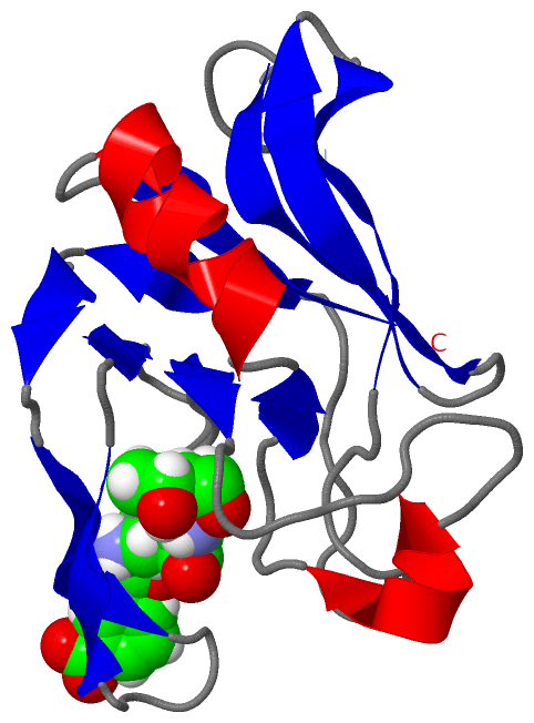
Ligands, Modified Residues, Ions (1, 1)
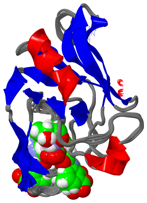
NMR Structure (1, 1)
|
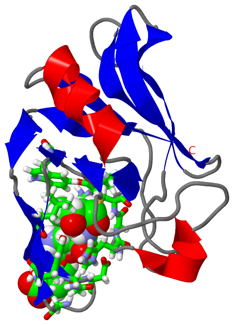
Sites (1, 1)
NMR Structure (1, 1)
|
SS Bonds (0, 0)| (no "SS Bond" information available for 3ZGP) |
Cis Peptide Bonds (0, 0)| (no "Cis Peptide Bond" information available for 3ZGP) |
SAPs(SNPs)/Variants (0, 0)| (no "SAP(SNP)/Variant" information available for 3ZGP) |
PROSITE Motifs (0, 0)| (no "PROSITE Motif" information available for 3ZGP) |
Exons (0, 0)| (no "Exon" information available for 3ZGP) |
Sequences/Alignments
NMR StructureChain A from PDB Type:PROTEIN Length:129 aligned with Q3Y185_ENTFC | Q3Y185 from UniProtKB/TrEMBL Length:466 Alignment length:135 341 351 361 371 381 391 401 411 421 431 441 451 461 Q3Y185_ENTFC 332 GTTADHPLIEDTYIEVDLENQHMWYYKDGKVALETDIVSGKPTTPTPAGVFYVWNKEEDATLKGTNDDGTPYESPVNYWMPIDWTGVGIHDSDWQPEYGGDLWKTRGSHGCINTPPSVMKELFGMVEKGTPVLVF 466 SCOP domains --------------------------------------------------------------------------------------------------------------------------------------- SCOP domains CATH domains --------------------------------------------------------------------------------------------------------------------------------------- CATH domains Pfam domains --------------------------------------------------------------------------------------------------------------------------------------- Pfam domains
|
||||||||||||||||||||
SCOP Domains (0, 0)| (no "SCOP Domain" information available for 3ZGP) |
CATH Domains (0, 0)| (no "CATH Domain" information available for 3ZGP) |
Pfam Domains (0, 0)| (no "Pfam Domain" information available for 3ZGP) |
Gene Ontology (3, 3)|
NMR Structure(hide GO term definitions) Chain A (Q3Y185_ENTFC | Q3Y185)
|
||||||||||||||||||||||||||||||
Interactive Views
|
||||||||||||||||||||||||||||||||||||||||||||||||||||||||||||||||||||||||||||||||||||||||||||||||||||||||||||||||||||||
Still Images
|
||||||||||||||||
Databases
|
||||||||||||||||||||||||||||||||||||||||||||||||||||||||||||||||||||||||||||||||||||||||||||||||||||||||||||||||||||||||||||||||||||||||||||||||||||||||||||||||
Analysis Tools
|
|||||||||||||||||||||||||||||||||||||||||||||||||||||||||||||
Entries Sharing at Least One Protein Chain (UniProt ID)
Related Entries Specified in the PDB File
|
|