Show PDB file:   
         Plain Text   HTML   (compressed file size)
QuickSearch:   
by PDB,NDB,UniProt,PROSITE Code or Search Term(s)  
(-)Biol.Unit 1 - manually
(-)Asymmetric Unit
(-)Asym. Unit - sites
(-)Biological Unit 1
(-)Biol. Unit 1 - sites
(-)Biological Unit 2
(-)Biological Unit 3
(-)Biological Unit 4
collapse expand < >
Image Biol.Unit 1 - manually
Biol.Unit 1 - manually  (Jmol Viewer)
Image Asymmetric Unit
Asymmetric Unit  (Jmol Viewer)
Image Asym. Unit - sites
Asym. Unit - sites  (Jmol Viewer)
Image Biological Unit 1
Biological Unit 1  (Jmol Viewer)
Image Biol. Unit 1 - sites
Biol. Unit 1 - sites  (Jmol Viewer)
Image Biological Unit 2
Biological Unit 2  (Jmol Viewer)
Image Biological Unit 3
Biological Unit 3  (Jmol Viewer)
Image Biological Unit 4
Biological Unit 4  (Jmol Viewer)

(-) Description

Title :  CRYSTAL STRUCTURE OF MOUSE TRNA(SEC)
 
Authors :  M. C. Wahl, O. M. Ganichkin, E. A. Anedchenko
Date :  07 Apr 11  (Deposition) - 08 Jun 11  (Release) - 11 Apr 12  (Revision)
Method :  X-RAY DIFFRACTION
Resolution :  2.00
Chains :  Asym. Unit :  A,B
Biol. Unit 1:  A  (1x)
Biol. Unit 2:  B  (1x)
Biol. Unit 3:  A,B  (1x)
Biol. Unit 4:  A (1x),B (1x)
Keywords :  Metal Binding By Rna, Rna Hydration, Structural Flexibility, Trnasec, Trna Fold, Translation, Serrs, Secs, Pstk, Rna (Keyword Search: [Gene Ontology, PubMed, Web (Google))
 
Reference :  O. M. Ganichkin, E. A. Anedchenko, M. C. Wahl
Crystal Structure Analysis Reveals Functional Flexibility I The Selenocysteine-Specific Trna From Mouse.
Plos One V. 6 20032 2011
PubMed-ID: 21629646  |  Reference-DOI: 10.1371/JOURNAL.PONE.0020032

(-) Compounds

Molecule 1 - TRNA(SEC)
    ChainsA, B
    EngineeredYES
    Organism CommonMOUSE
    Organism ScientificMUS MUSCULUS
    Organism Taxid10090
    Other DetailsRNA 86-MER
    Other Details - SourceSYNTHESIZED BY IN VITRO TRANSCRIPTION
    SyntheticYES

 Structural Features

(-) Chains, Units

  12
Asymmetric Unit AB
Biological Unit 1 (1x)A 
Biological Unit 2 (1x) B
Biological Unit 3 (1x)AB
Biological Unit 4 (1x)A (1x)B (1x)

Summary Information (see also Sequences/Alignments below)

(-) Ligands, Modified Residues, Ions  (3, 6)

Asymmetric Unit (3, 6)
No.NameCountTypeFull Name
1ACT2Ligand/IonACETATE ION
2GOL2Ligand/IonGLYCEROL
3SO42Ligand/IonSULFATE ION
Biological Unit 1 (3, 3)
No.NameCountTypeFull Name
1ACT1Ligand/IonACETATE ION
2GOL1Ligand/IonGLYCEROL
3SO41Ligand/IonSULFATE ION
Biological Unit 2 (3, 3)
No.NameCountTypeFull Name
1ACT1Ligand/IonACETATE ION
2GOL1Ligand/IonGLYCEROL
3SO41Ligand/IonSULFATE ION
Biological Unit 3 (3, 6)
No.NameCountTypeFull Name
1ACT2Ligand/IonACETATE ION
2GOL2Ligand/IonGLYCEROL
3SO42Ligand/IonSULFATE ION
Biological Unit 4 (3, 3)
No.NameCountTypeFull Name
1ACT1Ligand/IonACETATE ION
2GOL1Ligand/IonGLYCEROL
3SO41Ligand/IonSULFATE ION

(-) Sites  (6, 6)

Asymmetric Unit (6, 6)
No.NameEvidenceResiduesDescription
1AC1SOFTWAREC A:10 , C A:11 , HOH A:118 , HOH A:142 , HOH A:157BINDING SITE FOR RESIDUE SO4 A 73
2AC2SOFTWAREC B:10 , C B:11 , HOH B:168BINDING SITE FOR RESIDUE SO4 B 73
3AC3SOFTWAREC A:10 , U A:26 , G A:27 , HOH A:129 , G B:25 , ACT B:75 , HOH B:79BINDING SITE FOR RESIDUE GOL A 74
4AC4SOFTWAREG A:30 , G A:31 , HOH A:175 , HOH A:191 , G B:30 , G B:31 , U B:41 , HOH B:193 , HOH B:195BINDING SITE FOR RESIDUE GOL B 74
5AC5SOFTWAREC A:11 , GOL A:74 , HOH A:129 , C B:10 , U B:26 , G B:27 , HOH B:139BINDING SITE FOR RESIDUE ACT B 75
6AC6SOFTWAREG A:22BINDING SITE FOR RESIDUE ACT A 75

(-) SS Bonds  (0, 0)

(no "SS Bond" information available for 3RG5)

(-) Cis Peptide Bonds  (0, 0)

(no "Cis Peptide Bond" information available for 3RG5)

 Sequence-Structure Mapping

(-) SAPs(SNPs)/Variants  (0, 0)

(no "SAP(SNP)/Variant" information available for 3RG5)

(-) PROSITE Motifs  (0, 0)

(no "PROSITE Motif" information available for 3RG5)

(-) Exons   (0, 0)

(no "Exon" information available for 3RG5)

(-) Sequences/Alignments

Asymmetric Unit
   Reformat: Number of residues per line =  ('0' or empty: single-line sequence representation)
  Number of residues per labelling interval =   
  UniProt sequence: complete  aligned part    
   Show mapping: SCOP domains CATH domains Pfam domains Secondary structure (by author)
SAPs(SNPs) PROSITE motifs Exons
(details for a mapped element are shown in a popup box when the mouse pointer rests over it)
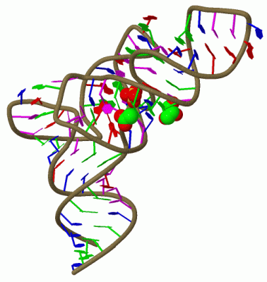
Chain A from PDB  Type:RNA  Length:86
                                                                                                                      
                 3rg5 A   1 GCCCGGAUGAUCCUCAGUGGUCUGGGGUGCAGGCUUCAAACCUGUAGCUGUUUAGCGACAGAGUGGUUCAAUUCCACCUUUCGGGC  72
                                 ||  8       |19 |      28        38       47A|||||||47K|||     57      |67A|     
                                5A|         16|  |                         47A||||||47J||||            64| ||     
                                 5B          18  |                          47B||||||47K|||             66 ||     
                                               20A                           47C||||||47L||              67A|     
                                                                              47D|||||  48|               67B     
                                                                               47E||||   50                       
                                                                                47F|||                            
                                                                                 47G||                            
                                                                                  47H|                            
                                                                                   47I                            

Chain B from PDB  Type:RNA  Length:86
                                                                                                                      
                 3rg5 B   1 GCCCGGAUGAUCCUCAGUGGUCUGGGGUGCAGGCUUCAAACCUGUAGCUGUUUAGCGACAGAGUGGUUCAAUUCCACCUUUCGGGC  72
                                 ||  8       |19 |      28        38       47A|||||||47K|||     57      |67A|     
                                5A|         16|  |                         47A||||||47J||||            64| ||     
                                 5B          18  |                          47B||||||47K|||             66 ||     
                                               20A                           47C||||||47L||              67A|     
                                                                              47D|||||  48|               67B     
                                                                               47E||||   50                       
                                                                                47F|||                            
                                                                                 47G||                            
                                                                                  47H|                            
                                                                                   47I                            

   Legend:   → Mismatch (orange background)
  - → Gap (green background, '-', border residues have a numbering label)
    → Modified Residue (blue background, lower-case, 'x' indicates undefined single-letter code, labelled with number + name)
  x → Chemical Group (purple background, 'x', labelled with number + name, e.g. ACE or NH2)
  extra numbering lines below/above indicate numbering irregularities and modified residue names etc., number ends below/above '|'

 Classification and Annotation

(-) SCOP Domains  (0, 0)

(no "SCOP Domain" information available for 3RG5)

(-) CATH Domains  (0, 0)

(no "CATH Domain" information available for 3RG5)

(-) Pfam Domains  (0, 0)

(no "Pfam Domain" information available for 3RG5)

(-) Gene Ontology  (0, 0)

Asymmetric Unit(hide GO term definitions)
    (no "Gene Ontology" information available for 3RG5)

 Visualization

(-) Interactive Views

Asymmetric Unit
  Complete Structure
    Jena3D(integrated viewing of ligand, site, SAP, PROSITE, SCOP information)
    WebMol | AstexViewer[tm]@PDBe
(Java Applets, require no local installation except for Java; loading may be slow)
    STRAP
(Java WebStart application, automatic local installation, requires Java; full application with system access!)
    RasMol
(require local installation)
    Molscript (VRML)
(requires installation of a VRML viewer; select preferred view via VRML and generate a mono or stereo PDF format file)
 
  Ligands, Modified Residues, Ions
    ACT  [ RasMol | Jena3D ]  +environment [ RasMol | Jena3D ]
    GOL  [ RasMol | Jena3D ]  +environment [ RasMol | Jena3D ]
    SO4  [ RasMol | Jena3D ]  +environment [ RasMol | Jena3D ]
 
  Sites
    AC1  [ RasMol ]  +environment [ RasMol ]
    AC2  [ RasMol ]  +environment [ RasMol ]
    AC3  [ RasMol ]  +environment [ RasMol ]
    AC4  [ RasMol ]  +environment [ RasMol ]
    AC5  [ RasMol ]  +environment [ RasMol ]
    AC6  [ RasMol ]  +environment [ RasMol ]
 
  Cis Peptide Bonds
(no "Cis Peptide Bonds" information available for 3rg5)
 
Biological Units
  Complete Structure
    Biological Unit 1  [ Jena3D ]
    Biological Unit 2  [ Jena3D ]
    Biological Unit 3  [ Jena3D ]
    Biological Unit 4  [ Jena3D ]

(-) Still Images

Jmol
  protein: cartoon or spacefill or dots and stick; nucleic acid: cartoon and stick; ligands: spacefill; active site: stick
Molscript
  protein, nucleic acid: cartoon; ligands: spacefill; active site: ball and stick

 Databases and Analysis Tools

(-) Databases

Access by PDB/NDB ID
  3rg5
    Family and Domain InformationProDom | SYSTERS
    General Structural InformationGlycoscienceDB | MMDB | NDB | OCA | PDB | PDBe | PDBj | PDBsum | PDBWiki | PQS | PROTEOPEDIA
    Orientation in MembranesOPM
    Protein SurfaceSURFACE
    Secondary StructureDSSP | HSSP
    Structural GenomicsGeneCensus
    Structural NeighboursCE | VAST
    Structure ClassificationCATH | Dali | SCOP
    Validation and Original DataBMRB Data View | BMRB Restraints Grid | EDS | PROCHECK | RECOORD | WHAT_CHECK
 
Access by UniProt ID/Accession number
  (no 'UniProt ID/Accession number' available) |
    Comparative Protein Structure ModelsModBase
    Genomic InformationEnsembl
    Protein-protein InteractionDIP
    Sequence, Family and Domain InformationInterPro | Pfam | SMART | UniProt
 
Access by Enzyme Classificator   (EC Number)
  (no 'Enzyme Classificator' available)
    General Enzyme InformationBRENDA | EC-PDB | Enzyme | IntEnz
    PathwayKEGG | MetaCyc
 
Access by Disease Identifier   (MIM ID)
  (no 'MIM ID' available)
    Disease InformationOMIM
 
Access by GenAge ID
  (no 'GenAge ID' available)
    Age Related InformationGenAge

(-) Analysis Tools

Access by PDB/NDB ID
    Domain InformationXDom
    Interatomic Contacts of Structural UnitsCSU
    Ligand-protein ContactsLPC
    Protein CavitiescastP
    Sequence and Secondary StructurePDBCartoon
    Structure AlignmentSTRAP(Java WebStart application, automatic local installation, requires Java; full application with system access!)
    Structure and Sequence BrowserSTING
 
Access by UniProt ID/Accession number
  (no 'UniProt ID/Accession number' available) |
    Protein Disorder PredictionDisEMBL | FoldIndex | GLOBPLOT

 Related Entries

(-) Entries Sharing at Least One Protein Chain (UniProt ID)

(no "Entries Sharing at Least One Protein Chain" available for 3RG5)

(-) Related Entries Specified in the PDB File

(no "Related Entries Specified in the PDB File" available for 3RG5)