| molecular function |
|---|
| | GO:0005524 | | ATP binding | | Interacting selectively and non-covalently with ATP, adenosine 5'-triphosphate, a universally important coenzyme and enzyme regulator. |
| | GO:0051015 | | actin filament binding | | Interacting selectively and non-covalently with an actin filament, also known as F-actin, a helical filamentous polymer of globular G-actin subunits. |
| | GO:0042805 | | actinin binding | | Interacting selectively and non-covalently with actinin, any member of a family of proteins that crosslink F-actin. |
| | GO:0005509 | | calcium ion binding | | Interacting selectively and non-covalently with calcium ions (Ca2+). |
| | GO:0005516 | | calmodulin binding | | Interacting selectively and non-covalently with calmodulin, a calcium-binding protein with many roles, both in the calcium-bound and calcium-free states. |
| | GO:0019899 | | enzyme binding | | Interacting selectively and non-covalently with any enzyme. |
| | GO:0042802 | | identical protein binding | | Interacting selectively and non-covalently with an identical protein or proteins. |
| | GO:0016301 | | kinase activity | | Catalysis of the transfer of a phosphate group, usually from ATP, to a substrate molecule. |
| | GO:0046872 | | metal ion binding | | Interacting selectively and non-covalently with any metal ion. |
| | GO:0051371 | | muscle alpha-actinin binding | | Interacting selectively and non-covalently with muscle isoforms of actinin. Muscle alpha-actinin isoforms are found in skeletal and cardiac muscle and are localized to the Z-disc. |
| | GO:0000166 | | nucleotide binding | | Interacting selectively and non-covalently with a nucleotide, any compound consisting of a nucleoside that is esterified with (ortho)phosphate or an oligophosphate at any hydroxyl group on the ribose or deoxyribose. |
| | GO:0002020 | | protease binding | | Interacting selectively and non-covalently with any protease or peptidase. |
| | GO:0005515 | | protein binding | | Interacting selectively and non-covalently with any protein or protein complex (a complex of two or more proteins that may include other nonprotein molecules). |
| | GO:0004672 | | protein kinase activity | | Catalysis of the phosphorylation of an amino acid residue in a protein, usually according to the reaction: a protein + ATP = a phosphoprotein + ADP. |
| | GO:0019901 | | protein kinase binding | | Interacting selectively and non-covalently with a protein kinase, any enzyme that catalyzes the transfer of a phosphate group, usually from ATP, to a protein substrate. |
| | GO:0043621 | | protein self-association | | Interacting selectively and non-covalently with a domain within the same polypeptide. |
| | GO:0004674 | | protein serine/threonine kinase activity | | Catalysis of the reactions: ATP + protein serine = ADP + protein serine phosphate, and ATP + protein threonine = ADP + protein threonine phosphate. |
| | GO:0004713 | | protein tyrosine kinase activity | | Catalysis of the reaction: ATP + a protein tyrosine = ADP + protein tyrosine phosphate. |
| | GO:0008307 | | structural constituent of muscle | | The action of a molecule that contributes to the structural integrity of a muscle fiber. |
| | GO:0097493 | | structural molecule activity conferring elasticity | | The action of a molecule that contributes to the structural integrity of a complex or assembly within or outside a cell, providing elasticity and recoiling. |
| | GO:0031433 | | telethonin binding | | Interacting selectively and non-covalently with telethonin, a protein found in the Z disc of striated muscle and which is a substrate of the titin kinase. |
| | GO:0016740 | | transferase activity | | Catalysis of the transfer of a group, e.g. a methyl group, glycosyl group, acyl group, phosphorus-containing, or other groups, from one compound (generally regarded as the donor) to another compound (generally regarded as the acceptor). Transferase is the systematic name for any enzyme of EC class 2. |
| biological process |
|---|
| | GO:0060048 | | cardiac muscle contraction | | Muscle contraction of cardiac muscle tissue. |
| | GO:0048739 | | cardiac muscle fiber development | | The process whose specific outcome is the progression of cardiac muscle fiber over time, from its formation to the mature structure. |
| | GO:0003300 | | cardiac muscle hypertrophy | | The enlargement or overgrowth of all or part of the heart muscle due to an increase in size of cardiac muscle cells without cell division. |
| | GO:0055008 | | cardiac muscle tissue morphogenesis | | The process in which the anatomical structures of cardiac muscle tissue are generated and organized. |
| | GO:0055003 | | cardiac myofibril assembly | | The process whose specific outcome is the progression of the cardiac myofibril over time, from its formation to the mature structure. A cardiac myofibril is a myofibril specific to cardiac muscle cells. |
| | GO:0035995 | | detection of muscle stretch | | The series of events by which a muscle stretch stimulus is received by a cell and converted into a molecular signal. |
| | GO:0007076 | | mitotic chromosome condensation | | The cell cycle process in which chromatin structure is compacted prior to and during mitosis in eukaryotic cells. |
| | GO:0006936 | | muscle contraction | | A process in which force is generated within muscle tissue, resulting in a change in muscle geometry. Force generation involves a chemo-mechanical energy conversion step that is carried out by the actin/myosin complex activity, which generates force through ATP hydrolysis. |
| | GO:0030049 | | muscle filament sliding | | The sliding of actin thin filaments and myosin thick filaments past each other in muscle contraction. This involves a process of interaction of myosin located on a thick filament with actin located on a thin filament. During this process ATP is split and forces are generated. |
| | GO:0018108 | | peptidyl-tyrosine phosphorylation | | The phosphorylation of peptidyl-tyrosine to form peptidyl-O4'-phospho-L-tyrosine. |
| | GO:0016310 | | phosphorylation | | The process of introducing a phosphate group into a molecule, usually with the formation of a phosphoric ester, a phosphoric anhydride or a phosphoric amide. |
| | GO:0002576 | | platelet degranulation | | The regulated exocytosis of secretory granules containing preformed mediators such as histamine and serotonin by a platelet. |
| | GO:0006468 | | protein phosphorylation | | The process of introducing a phosphate group on to a protein. |
| | GO:0050790 | | regulation of catalytic activity | | Any process that modulates the activity of an enzyme. |
| | GO:0045859 | | regulation of protein kinase activity | | Any process that modulates the frequency, rate or extent of protein kinase activity. |
| | GO:0051592 | | response to calcium ion | | Any process that results in a change in state or activity of a cell or an organism (in terms of movement, secretion, enzyme production, gene expression, etc.) as a result of a calcium ion stimulus. |
| | GO:0045214 | | sarcomere organization | | The myofibril assembly process that results in the organization of muscle actomyosin into sarcomeres. The sarcomere is the repeating unit of a myofibril in a muscle cell, composed of an array of overlapping thick and thin filaments between two adjacent Z discs. |
| | GO:0048769 | | sarcomerogenesis | | The process in which sarcomeres are added in series within a fiber. |
| | GO:0030241 | | skeletal muscle myosin thick filament assembly | | The aggregation, arrangement and bonding together of proteins to form the myosin-based thick filaments of myofibrils in skeletal muscle. |
| | GO:0030240 | | skeletal muscle thin filament assembly | | The aggregation, arrangement and bonding together of proteins to form the actin-based thin filaments of myofibrils in skeletal muscle. |
| | GO:0006941 | | striated muscle contraction | | A process in which force is generated within striated muscle tissue, resulting in the shortening of the muscle. Force generation involves a chemo-mechanical energy conversion step that is carried out by the actin/myosin complex activity, which generates force through ATP hydrolysis. Striated muscle is a type of muscle in which the repeating units (sarcomeres) of the contractile myofibrils are arranged in registry throughout the cell, resulting in transverse or oblique striations observable at the level of the light microscope. |
| cellular component |
|---|
| | GO:0031674 | | I band | | A region of a sarcomere that appears as a light band on each side of the Z disc, comprising a region of the sarcomere where thin (actin) filaments are not overlapped by thick (myosin) filaments; contains actin, troponin, and tropomyosin; each sarcomere includes half of an I band at each end. |
| | GO:0031430 | | M band | | The midline of aligned thick filaments in a sarcomere; location of specific proteins that link thick filaments. Depending on muscle type the M band consists of different numbers of M lines. |
| | GO:0030018 | | Z disc | | Platelike region of a muscle sarcomere to which the plus ends of actin filaments are attached. |
| | GO:0000794 | | condensed nuclear chromosome | | A highly compacted molecule of DNA and associated proteins resulting in a cytologically distinct nuclear chromosome. |
| | GO:0005737 | | cytoplasm | | All of the contents of a cell excluding the plasma membrane and nucleus, but including other subcellular structures. |
| | GO:0005829 | | cytosol | | The part of the cytoplasm that does not contain organelles but which does contain other particulate matter, such as protein complexes. |
| | GO:0070062 | | extracellular exosome | | A vesicle that is released into the extracellular region by fusion of the limiting endosomal membrane of a multivesicular body with the plasma membrane. Extracellular exosomes, also simply called exosomes, have a diameter of about 40-100 nm. |
| | GO:0005576 | | extracellular region | | The space external to the outermost structure of a cell. For cells without external protective or external encapsulating structures this refers to space outside of the plasma membrane. This term covers the host cell environment outside an intracellular parasite. |
| | GO:0005859 | | muscle myosin complex | | A filament of myosin found in a muscle cell of any type. |
| | GO:0005634 | | nucleus | | A membrane-bounded organelle of eukaryotic cells in which chromosomes are housed and replicated. In most cells, the nucleus contains all of the cell's chromosomes except the organellar chromosomes, and is the site of RNA synthesis and processing. In some species, or in specialized cell types, RNA metabolism or DNA replication may be absent. |
| | GO:0005865 | | striated muscle thin filament | | Filaments formed of actin and associated proteins; attached to Z discs at either end of sarcomeres in myofibrils. |
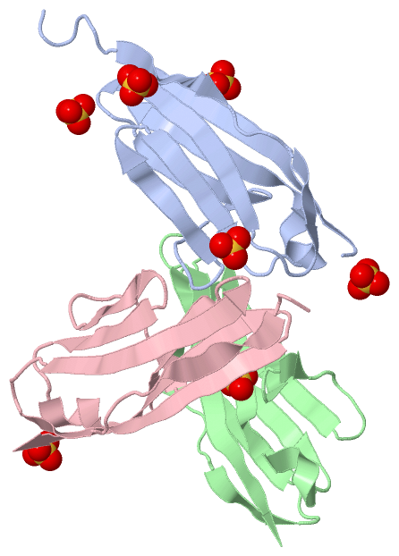
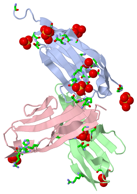
 Description
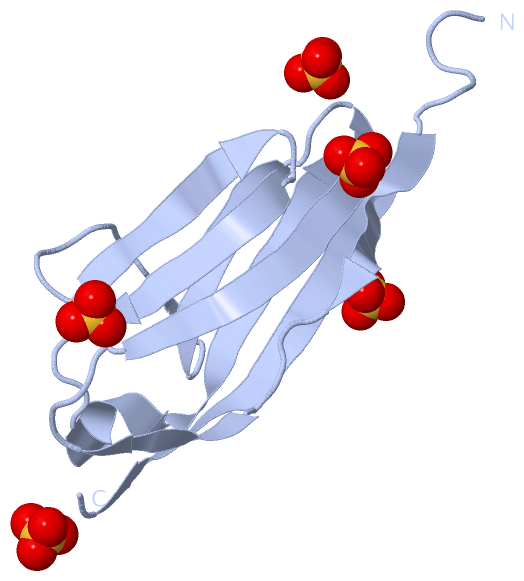
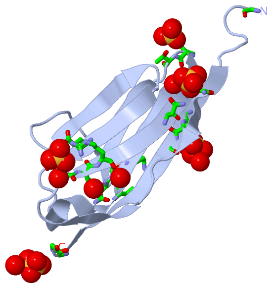