|
|
|
|
 Description
|
|
Compounds
|
||||||||||||||||||||||||||||||||||||||||||||||||
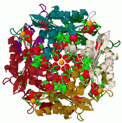
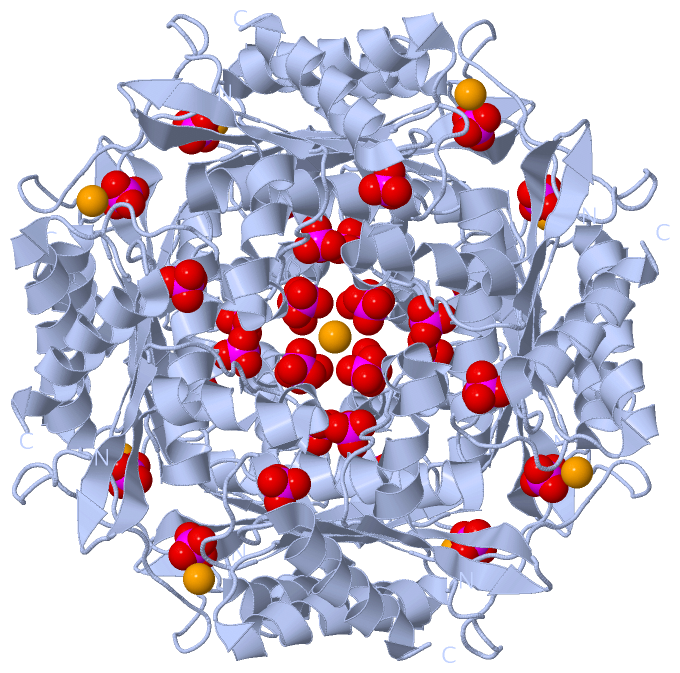
Chains, Units
Summary Information (see also Sequences/Alignments below) |
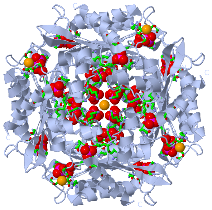
Ligands, Modified Residues, Ions (2, 6)| Asymmetric Unit (2, 6) Biological Unit 1 (1, 32) |
Sites (6, 6)
Asymmetric Unit (6, 6)
|
SS Bonds (0, 0)| (no "SS Bond" information available for 3FPW) |
Cis Peptide Bonds (1, 1)
Asymmetric Unit
|
||||||||
SAPs(SNPs)/Variants (0, 0)| (no "SAP(SNP)/Variant" information available for 3FPW) |
PROSITE Motifs (0, 0)| (no "PROSITE Motif" information available for 3FPW) |
Exons (0, 0)| (no "Exon" information available for 3FPW) |
Sequences/Alignments
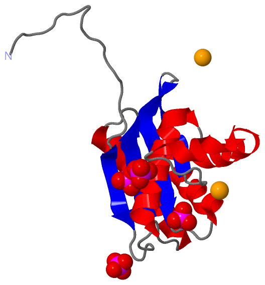
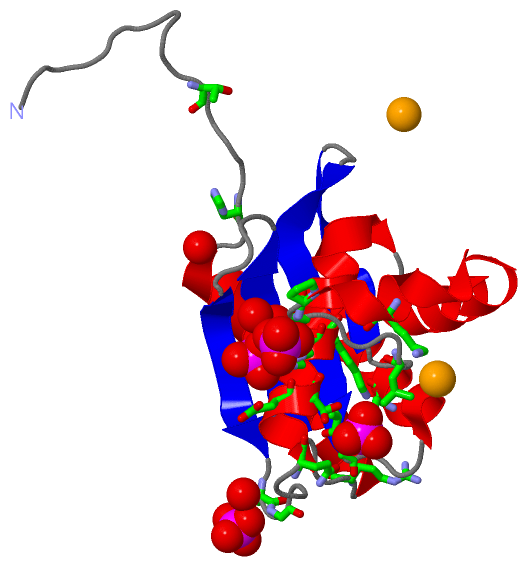
Asymmetric UnitChain A from PDB Type:PROTEIN Length:140 aligned with Q9RIM2_STRRE | Q9RIM2 from UniProtKB/TrEMBL Length:188 Alignment length:140 56 66 76 86 96 106 116 126 136 146 156 166 176 186 Q9RIM2_STRRE 47 PVAARGGELTQSTHLTLEAATKAARAAVEAAEKDGRHVSVAVVDRNGNTLVTLRGDGAGPQSYESAERKAFTAVSWNAPTSELAKRLAQAPTLKDIPGTLFLAGGTPVTAKGAPVAGIGVAGAPSGDLDEQYARAGAAVL 186 SCOP domains -------------------------------------------------------------------------------------------------------------------------------------------- SCOP domains CATH domains -------------------------------------------------------------------------------------------------------------------------------------------- CATH domains Pfam domains -------------------------------------------------------------------------------------------------------------------------------------------- Pfam domains SAPs(SNPs) -------------------------------------------------------------------------------------------------------------------------------------------- SAPs(SNPs) PROSITE -------------------------------------------------------------------------------------------------------------------------------------------- PROSITE Transcript -------------------------------------------------------------------------------------------------------------------------------------------- Transcript 3fpw A 15 PVAARGGELTQSTHLTLEAATKAARAAVEAAEKDGRHVSVAVVDRNGNTLVTLRGDGAGPQSYESAERKAFTAVSWNAPTSELAKRLAQAPTLKDIPGTLFLAGGTPVTAKGAPVAGIGVAGAPSGDLDEQYARAGAAVL 154 24 34 44 54 64 74 84 94 104 114 124 134 144 154
|
||||||||||||||||||||
SCOP Domains (0, 0)| (no "SCOP Domain" information available for 3FPW) |
CATH Domains (0, 0)| (no "CATH Domain" information available for 3FPW) |
Pfam Domains (0, 0)| (no "Pfam Domain" information available for 3FPW) |
Gene Ontology (0, 0)|
Asymmetric Unit(hide GO term definitions)
(no "Gene Ontology" information available for 3FPW)
|
Interactive Views
|
|||||||||||||||||||||||||||||||||||||||||||||||||||||||||||||||||||||||||||||||||||||||||||||||||||||||||||||||||||||||||||||||||||||||||||||||||||||||||||||||||||||||||||||||||||
Still Images
|
||||||||||||||||
Databases
|
||||||||||||||||||||||||||||||||||||||||||||||||||||||||||||||||||||||||||||||||||||||||||||||||||||||||||||||||||||||||||||||||||||||||||||||||||||||||||||||||
Analysis Tools
|
|||||||||||||||||||||||||||||||||||||||||||||||||||||||||||||
Entries Sharing at Least One Protein Chain (UniProt ID)
Related Entries Specified in the PDB File
|
|