|
|
|
|
 Description
|
|
Compounds
|
||||||||||||||||||||||||||||||||||||||||||||||||
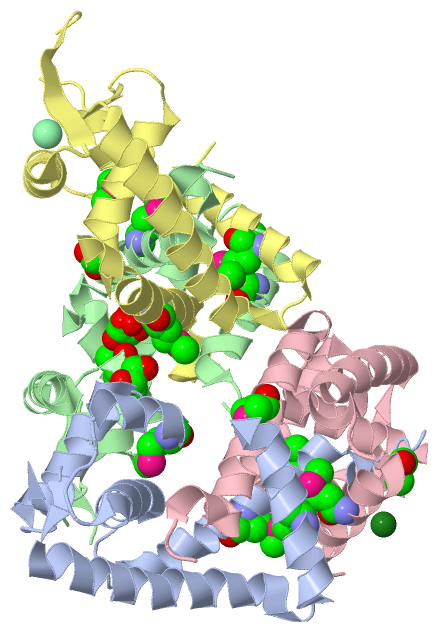
Chains, Units
Summary Information (see also Sequences/Alignments below) |
Ligands, Modified Residues, Ions (5, 19)
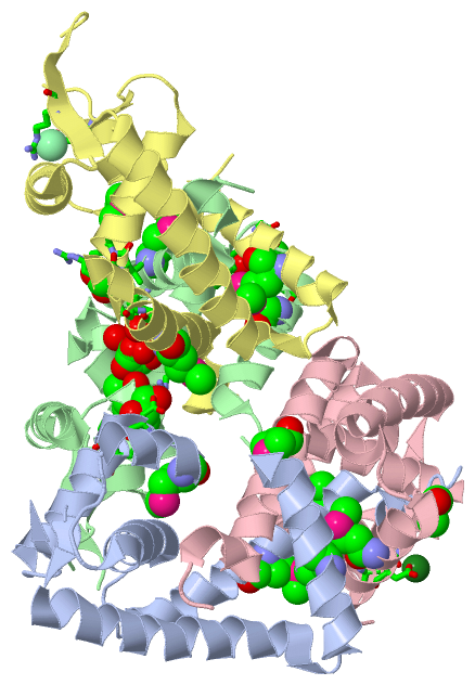
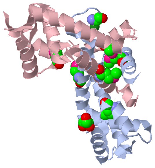
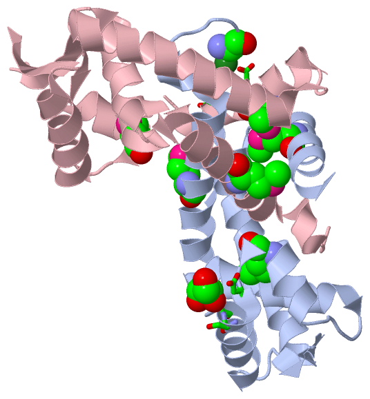
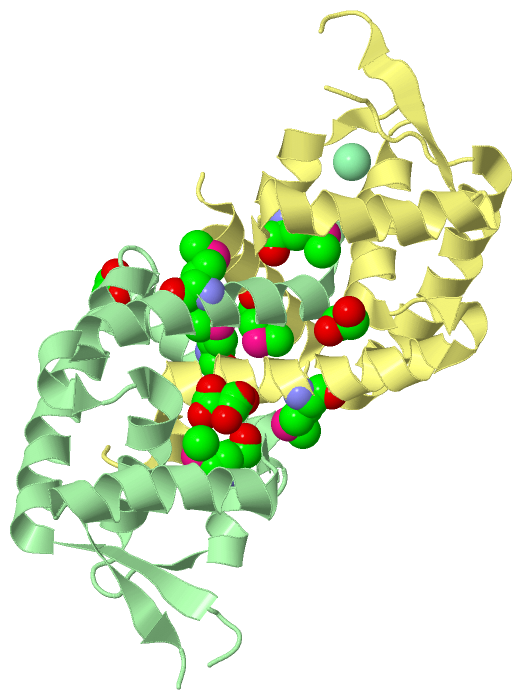
Asymmetric Unit (5, 19)
|
Sites (6, 6)
Asymmetric Unit (6, 6)
|
SS Bonds (0, 0)| (no "SS Bond" information available for 3FM5) |
Cis Peptide Bonds (0, 0)| (no "Cis Peptide Bond" information available for 3FM5) |
SAPs(SNPs)/Variants (0, 0)| (no "SAP(SNP)/Variant" information available for 3FM5) |
PROSITE Motifs (0, 0)| (no "PROSITE Motif" information available for 3FM5) |
Exons (0, 0)| (no "Exon" information available for 3FM5) |
Sequences/Alignments
Asymmetric UnitChain A from PDB Type:PROTEIN Length:131 aligned with Q0S6D0_RHOJR | Q0S6D0 from UniProtKB/TrEMBL Length:146 Alignment length:142 10 20 30 40 50 60 70 80 90 100 110 120 130 140 Q0S6D0_RHOJR 1 MAESQALSDDIGFLLSRVGGMVLGAVNKALVPTGLRVRSYSVLVLACEQAEGVNQRGVAATMGLDPSQIVGLVDELEERGLVVRTLDPSDRRNKLIAATEEGRRLRDDAKARVDAAHGRYFEGIPDTVVNQMRDTLQSIAFP 142 SCOP domains ---------------------------------------------------------------------------------------------------------------------------------------------- SCOP domains CATH domains -3fm5A00 A:2-142 'winged helix' repressor DNA binding domain CATH domains Pfam domains ---------------------------------------------------------------------------------------------------------------------------------------------- Pfam domains SAPs(SNPs) ---------------------------------------------------------------------------------------------------------------------------------------------- SAPs(SNPs) PROSITE ---------------------------------------------------------------------------------------------------------------------------------------------- PROSITE Transcript ---------------------------------------------------------------------------------------------------------------------------------------------- Transcript 3fm5 A 1 mAESQALSDDIGFLLSRVGGmVLGAVNKALVPTGLRVRSYSVLVLACEQAEGVNQRGVAATmGLDPSQIVGLVDELEERGLVVR-----------IAATEEGRRLRDDAKARVDAAHGRYFEGIPDTVVNQmRDTLQSIAFP 142 | 10 20| 30 40 50 60 | 70 80 | - | 100 110 120 130 | 140 | 21-MSE 62-MSE 84 96 132-MSE 1-MSE Chain B from PDB Type:PROTEIN Length:140 aligned with Q0S6D0_RHOJR | Q0S6D0 from UniProtKB/TrEMBL Length:146 Alignment length:142 13 23 33 43 53 63 73 83 93 103 113 123 133 143 Q0S6D0_RHOJR 4 SQALSDDIGFLLSRVGGMVLGAVNKALVPTGLRVRSYSVLVLACEQAEGVNQRGVAATMGLDPSQIVGLVDELEERGLVVRTLDPSDRRNKLIAATEEGRRLRDDAKARVDAAHGRYFEGIPDTVVNQMRDTLQSIAFPTFV 145 SCOP domains ---------------------------------------------------------------------------------------------------------------------------------------------- SCOP domains CATH domains 3fm5B00 B:4-145 'winged helix' repressor DNA binding domain CATH domains Pfam domains ---------------------------------------------------------------------------------------------------------------------------------------------- Pfam domains SAPs(SNPs) ---------------------------------------------------------------------------------------------------------------------------------------------- SAPs(SNPs) PROSITE ---------------------------------------------------------------------------------------------------------------------------------------------- PROSITE Transcript ---------------------------------------------------------------------------------------------------------------------------------------------- Transcript 3fm5 B 4 SQALSDDIGFLLSRVGGmVLGAVNKALVPTGLRVRSYSVLVLACEQAEGVNQRGVAATmGLDPSQIVGLVDELEERGLVVRTLDP--RRNKLIAATEEGRRLRDDAKARVDAAHGRYFEGIPDTVVNQmRDTLQSIAFPTFV 145 13 |23 33 43 53 63 73 83 | |93 103 113 123 133 143 21-MSE 62-MSE 88 91 132-MSE Chain C from PDB Type:PROTEIN Length:137 aligned with Q0S6D0_RHOJR | Q0S6D0 from UniProtKB/TrEMBL Length:146 Alignment length:140 13 23 33 43 53 63 73 83 93 103 113 123 133 143 Q0S6D0_RHOJR 4 SQALSDDIGFLLSRVGGMVLGAVNKALVPTGLRVRSYSVLVLACEQAEGVNQRGVAATMGLDPSQIVGLVDELEERGLVVRTLDPSDRRNKLIAATEEGRRLRDDAKARVDAAHGRYFEGIPDTVVNQMRDTLQSIAFPT 143 SCOP domains -------------------------------------------------------------------------------------------------------------------------------------------- SCOP domains CATH domains 3fm5C00 C:4-143 'winged helix' repressor DNA binding domain CATH domains Pfam domains -------------------------------------------------------------------------------------------------------------------------------------------- Pfam domains SAPs(SNPs) -------------------------------------------------------------------------------------------------------------------------------------------- SAPs(SNPs) PROSITE -------------------------------------------------------------------------------------------------------------------------------------------- PROSITE Transcript -------------------------------------------------------------------------------------------------------------------------------------------- Transcript 3fm5 C 4 SQALSDDIGFLLSRVGGmVLGAVNKALVPTGLRVRSYSVLVLACEQAEGVNQRGVAATmGLDPSQIVGLVDELEERGLVVRTLDP---RNKLIAATEEGRRLRDDAKARVDAAHGRYFEGIPDTVVNQmRDTLQSIAFPT 143 13 |23 33 43 53 63 73 83 | 93 103 113 123 133 143 21-MSE 62-MSE 88 92 132-MSE Chain D from PDB Type:PROTEIN Length:138 aligned with Q0S6D0_RHOJR | Q0S6D0 from UniProtKB/TrEMBL Length:146 Alignment length:140 13 23 33 43 53 63 73 83 93 103 113 123 133 143 Q0S6D0_RHOJR 4 SQALSDDIGFLLSRVGGMVLGAVNKALVPTGLRVRSYSVLVLACEQAEGVNQRGVAATMGLDPSQIVGLVDELEERGLVVRTLDPSDRRNKLIAATEEGRRLRDDAKARVDAAHGRYFEGIPDTVVNQMRDTLQSIAFPT 143 SCOP domains -------------------------------------------------------------------------------------------------------------------------------------------- SCOP domains CATH domains 3fm5D00 D:4-143 'winged helix' repressor DNA binding domain CATH domains Pfam domains -------------------------------------------------------------------------------------------------------------------------------------------- Pfam domains SAPs(SNPs) -------------------------------------------------------------------------------------------------------------------------------------------- SAPs(SNPs) PROSITE -------------------------------------------------------------------------------------------------------------------------------------------- PROSITE Transcript -------------------------------------------------------------------------------------------------------------------------------------------- Transcript 3fm5 D 4 SQALSDDIGFLLSRVGGmVLGAVNKALVPTGLRVRSYSVLVLACEQAEGVNQRGVAATmGLDPSQIVGLVDELEERGLVVRTLDPSD--NKLIAATEEGRRLRDDAKARVDAAHGRYFEGIPDTVVNQmRDTLQSIAFPT 143 13 |23 33 43 53 63 73 83 | 93 103 113 123 133 143 21-MSE 62-MSE 90 93 132-MSE
|
||||||||||||||||||||
SCOP Domains (0, 0)| (no "SCOP Domain" information available for 3FM5) |
CATH Domains (1, 4)
Asymmetric Unit
 |
Pfam Domains (0, 0)| (no "Pfam Domain" information available for 3FM5) |
Gene Ontology (4, 4)|
Asymmetric Unit(hide GO term definitions) Chain A,B,C,D (Q0S6D0_RHOJR | Q0S6D0)
|
||||||||||||||||||||||||||||||||||||
Interactive Views
|
|||||||||||||||||||||||||||||||||||||||||||||||||||||||||||||||||||||||||||||||||||||||||||||||||||||||||||||||||||||||||||||||||||||||||||||||||||||||||||||||||||||||||||||||||||||||||||||||||||||||||||||||||
Still Images
|
||||||||||||||||
Databases
|
||||||||||||||||||||||||||||||||||||||||||||||||||||||||||||||||||||||||||||||||||||||||||||||||||||||||||||||||||||||||||||||||||||||||||||||||||||||||||||||||
Analysis Tools
|
|||||||||||||||||||||||||||||||||||||||||||||||||||||||||||||
Entries Sharing at Least One Protein Chain (UniProt ID)
Related Entries Specified in the PDB File
|
|