|
|
|
|
 Description
|
|
Compounds
|
||||||||||||||||||||||||||||||||||||||
Chains, Units
Summary Information (see also Sequences/Alignments below) |
Ligands, Modified Residues, Ions (0, 0)| (no "Ligand,Modified Residues,Ions" information available for 3PDM) |
Sites (0, 0)| (no "Site" information available for 3PDM) |
SS Bonds (0, 0)| (no "SS Bond" information available for 3PDM) |
Cis Peptide Bonds (0, 0)| (no "Cis Peptide Bond" information available for 3PDM) |
SAPs(SNPs)/Variants (0, 0)| (no "SAP(SNP)/Variant" information available for 3PDM) |
PROSITE Motifs (0, 0)| (no "PROSITE Motif" information available for 3PDM) |
Exons (0, 0)| (no "Exon" information available for 3PDM) |
Sequences/Alignments
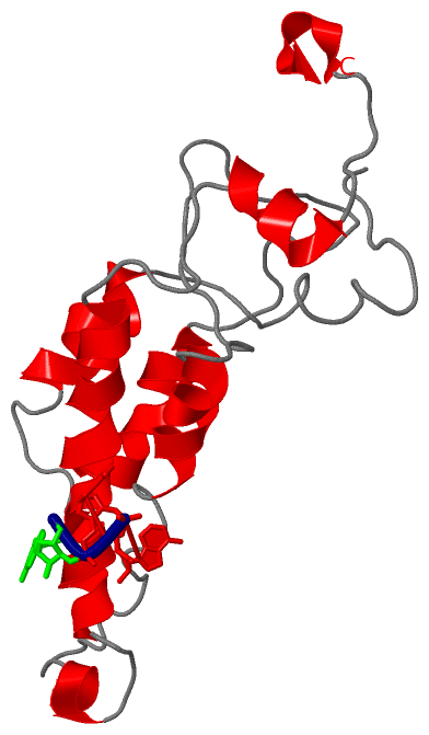
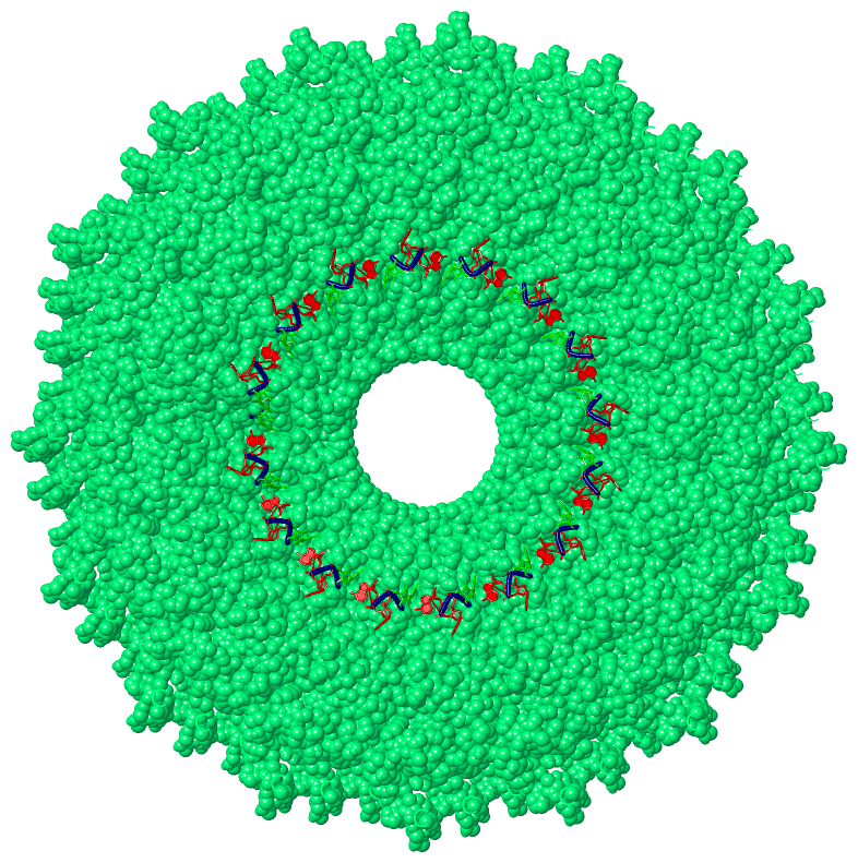
Asymmetric UnitChain P from PDB Type:PROTEIN Length:162 aligned with Q8BE68_9VIRU | Q8BE68 from UniProtKB/TrEMBL Length:163 Alignment length:162 11 21 31 41 51 61 71 81 91 101 111 121 131 141 151 161 Q8BE68_9VIRU 2 PYLNLTPLNLIYTSGTFAPYDVFLEILVKSRSNSFQTQAGRDTLREQLINSLQIVANLNTRYPLLGFYVWVRNPTLAPVFEALLRATDTKNRIIEVEEESRPTTAETLNATQRVDDATVAIHKEIDNILLLLQGGTAVYDRTAFEVVSGLSWADPTTTSTTT 163 SCOP domains ------------------------------------------------------------------------------------------------------------------------------------------------------------------ SCOP domains CATH domains ------------------------------------------------------------------------------------------------------------------------------------------------------------------ CATH domains Pfam domains TMV_coat-3pdmP01 P:1-145 ----------------- Pfam domains SAPs(SNPs) ------------------------------------------------------------------------------------------------------------------------------------------------------------------ SAPs(SNPs) PROSITE ------------------------------------------------------------------------------------------------------------------------------------------------------------------ PROSITE Transcript ------------------------------------------------------------------------------------------------------------------------------------------------------------------ Transcript 3pdm P 1 PYLNLTPLNLIYTSGTFAPYDVFLEILVKSRSNSFQTQAGRDTLREQLINSLQIVANLNTRYPLLGFYVWVRNPTLAPVFEALLRATDTKNRIIEVEEESRPTTAETLNATQRVDDATVAIHKEIDNILLLLQGGTAVYDRTAFEVASGLSWADPTTTSTTT 162 10 20 30 40 50 60 70 80 90 100 110 120 130 140 150 160
Chain R from PDB  Type:RNA  Length:3

                 3pdm R   1 GAA   3
|
||||||||||||||||||||
SCOP Domains (0, 0)| (no "SCOP Domain" information available for 3PDM) |
CATH Domains (0, 0)| (no "CATH Domain" information available for 3PDM) |
Pfam Domains (1, 1)
Asymmetric Unit
|
Gene Ontology (3, 3)|
Asymmetric Unit(hide GO term definitions) Chain P (Q8BE68_9VIRU | Q8BE68)
|
||||||||||||||||||||||||||||||
Interactive Views
|
||||||||||||||||||||||||||||||||||||||||||||||||||||||||||||||||||||||||||||||||||||||||||||||||||||||||||||||||||||||||||||||||||||||
Still Images
|
||||||||||||||||
Databases
|
||||||||||||||||||||||||||||||||||||||||||||||||||||||||||||||||||||||||||||||||||||||||||||||||||||||||||||||||||||||||||||||||||||||||||||||||||||||||||||||||
Analysis Tools
|
|||||||||||||||||||||||||||||||||||||||||||||||||||||||||||||
Entries Sharing at Least One Protein Chain (UniProt ID)
Related Entries Specified in the PDB File
|
|