Show PDB file:   
         Plain Text   HTML   (compressed file size)
QuickSearch:   
by PDB,NDB,UniProt,PROSITE Code or Search Term(s)  
(-)Asym./Biol. Unit
(-)Asym./Biol. Unit - sites
collapse expand < >
Image Asym./Biol. Unit
Asym./Biol. Unit  (Jmol Viewer)
Image Asym./Biol. Unit - sites
Asym./Biol. Unit - sites  (Jmol Viewer)

(-) Description

Title :  CRYSTAL STRUCTURE OF THE SMALL GTPASE RHEBL1
 
Authors :  L. Nedyalkova, N. Zhong, W. Tempel, Y. Tong, L. Shen, P. Loppnau, C. H. Ar A. M. Edwards, C. Bountra, J. Weigelt, A. Bochkarev, H. Park, Structur Genomics Consortium (Sgc)
Date :  13 Aug 10  (Deposition) - 25 Aug 10  (Release) - 25 Aug 10  (Revision)
Method :  X-RAY DIFFRACTION
Resolution :  2.30
Chains :  Asym./Biol. Unit :  A
Keywords :  Small Gtpase, Structural Genomics, Structural Genomics Consortium, Sgc, Hydrolase (Keyword Search: [Gene Ontology, PubMed, Web (Google))
 
Reference :  L. Nedyalkova, N. Zhong, W. Tempel, Y. Tong, L. Shen, P. Loppnau, C. H. Arrowsmith, A. M. Edwards, C. Bountra, J. Weigelt, A. Bochkarev, H. Park
Crystal Structure Of The Small Gtpase Rhebl1
To Be Published
PubMed: search

(-) Compounds

Molecule 1 - GTPASE RHEBL1
    ChainsA
    EngineeredYES
    Expression SystemESCHERICHIA COLI
    Expression System PlasmidPET28-MHL
    Expression System StrainBL21-V2R-PRARE2
    Expression System Taxid562
    Expression System Vector TypePLASMID
    GeneRHEBL1
    Organism CommonHUMAN
    Organism ScientificHOMO SAPIENS
    Organism Taxid9606
    SynonymRAS HOMOLOG ENRICHED IN BRAIN-LIKE PROTEIN 1, RHEB-LIKE PROTEIN 1, RHEB2, RAS HOMOLOG ENRICHED IN BRAIN LIKE-1 C, RHEBL1C

 Structural Features

(-) Chains, Units

  1
Asymmetric/Biological Unit A

Summary Information (see also Sequences/Alignments below)
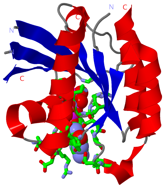
(-) Ligands, Modified Residues, Ions  (2, 2)

Asymmetric/Biological Unit (2, 2)
No.NameCountTypeFull Name
1GNP1Ligand/IonPHOSPHOAMINOPHOSPHONIC ACID-GUANYLATE ESTER
2MG1Ligand/IonMAGNESIUM ION

(-) Sites  (2, 2)

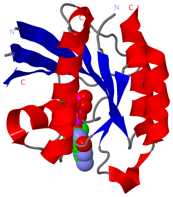
Asymmetric Unit (2, 2)
No.NameEvidenceResiduesDescription
1AC1SOFTWARETHR A:20 , THR A:38 , HOH A:184 , HOH A:185 , GNP A:201BINDING SITE FOR RESIDUE MG A 202
2AC2SOFTWAREARG A:15 , CYS A:16 , VAL A:17 , GLY A:18 , LYS A:19 , THR A:20 , SER A:21 , PHE A:31 , SER A:32 , GLU A:33 , GLY A:34 , TYR A:35 , PRO A:37 , THR A:38 , GLY A:63 , ASN A:119 , LYS A:120 , ASP A:122 , LEU A:123 , SER A:149 , ALA A:150 , ARG A:151 , LEU A:155 , HOH A:185 , HOH A:188 , MG A:202BINDING SITE FOR RESIDUE GNP A 201

(-) SS Bonds  (0, 0)

(no "SS Bond" information available for 3OES)

(-) Cis Peptide Bonds  (0, 0)

(no "Cis Peptide Bond" information available for 3OES)

 Sequence-Structure Mapping

(-) SAPs(SNPs)/Variants  (0, 0)

(no "SAP(SNP)/Variant" information available for 3OES)

(-) PROSITE Motifs  (0, 0)

(no "PROSITE Motif" information available for 3OES)

(-) Exons   (0, 0)

(no "Exon" information available for 3OES)

(-) Sequences/Alignments

Asymmetric/Biological Unit
   Reformat: Number of residues per line =  ('0' or empty: single-line sequence representation)
  Number of residues per labelling interval =   
  UniProt sequence: complete  aligned part    
   Show mapping: SCOP domains CATH domains Pfam domains Secondary structure (by author)
SAPs(SNPs) PROSITE motifs Exons
(details for a mapped element are shown in a popup box when the mouse pointer rests over it)
Chain A from PDB  Type:PROTEIN  Length:157
 aligned with REBL1_HUMAN | Q8TAI7 from UniProtKB/Swiss-Prot  Length:183

    Alignment length:166
                                    14        24        34        44        54        64        74        84        94       104       114       124       134       144       154       164      
          REBL1_HUMAN     5 RYRKVVILGYRCVGKTSLAHQFVEGEFSEGYDPTVENTYSKIVTLGKDEFHLHLVDTAGQDEYSILPYSFIIGVHGYVLVYSVTSLHSFQVIESLYQKLHEGHGKTRVPVVLVGNKADLSPEREVQAVEGKKLAESWGATFMESSARENQLTQGIFTKVIQEIARV 170
               SCOP domains d3oesa_ A: automated matches                                                                                                                                           SCOP domains
               CATH domains ---------------------------------------------------------------------------------------------------------------------------------------------------------------------- CATH domains
               Pfam domains ---Ras-3oesA01 A:8-169                                                                                                                                               - Pfam domains
         Sec.struct. author .eeeeeeee....hhhhhhhhhhhh..........eeeeeeee.----.eeeeeeee.........hhhhh....eeeeeee..hhhhhhhhhhhhhhhhh-----...eeeeee...hhhhh..hhhhhhhhhhhhh.eeee....hhhhhhhhhhhhhhhhhhh Sec.struct. author
                 SAPs(SNPs) ---------------------------------------------------------------------------------------------------------------------------------------------------------------------- SAPs(SNPs)
                    PROSITE ---------------------------------------------------------------------------------------------------------------------------------------------------------------------- PROSITE
                 Transcript ---------------------------------------------------------------------------------------------------------------------------------------------------------------------- Transcript
                 3oes A   5 RYRKVVILGYRCVGKTSLAHQFVEGEFSEGYDPTVENTYSKIVT----EFHLHLVDTAGQDEYSILPYSFIIGVHGYVLVYSVTSLHSFQVIESLYQKLHE-----RVPVVLVGNKADLSPEREVQAVEGKKLAESWGATFMESSARENQLTQGIFTKVIQEIARV 170
                                    14        24        34        44   |    54        64        74        84        94       104|     |114       124       134       144       154       164      
                                                                      48   53                                                 105   111                                                           

   Legend:   → Mismatch (orange background)
  - → Gap (green background, '-', border residues have a numbering label)
    → Modified Residue (blue background, lower-case, 'x' indicates undefined single-letter code, labelled with number + name)
  x → Chemical Group (purple background, 'x', labelled with number + name, e.g. ACE or NH2)
  extra numbering lines below/above indicate numbering irregularities and modified residue names etc., number ends below/above '|'

 Classification and Annotation

(-) SCOP Domains  (1, 1)

Asymmetric/Biological Unit

(-) CATH Domains  (0, 0)

(no "CATH Domain" information available for 3OES)

(-) Pfam Domains  (1, 1)

Asymmetric/Biological Unit
(-)
Family: Ras (176)

(-) Gene Ontology  (11, 11)

Asymmetric/Biological Unit(hide GO term definitions)
Chain A   (REBL1_HUMAN | Q8TAI7)
molecular function
    GO:0005525    GTP binding    Interacting selectively and non-covalently with GTP, guanosine triphosphate.
    GO:0046872    metal ion binding    Interacting selectively and non-covalently with any metal ion.
    GO:0000166    nucleotide binding    Interacting selectively and non-covalently with a nucleotide, any compound consisting of a nucleoside that is esterified with (ortho)phosphate or an oligophosphate at any hydroxyl group on the ribose or deoxyribose.
    GO:0005515    protein binding    Interacting selectively and non-covalently with any protein or protein complex (a complex of two or more proteins that may include other nonprotein molecules).
biological process
    GO:0031929    TOR signaling    A series of molecular signals mediated by TOR (Target of rapamycin) proteins, members of the phosphoinositide (PI) 3-kinase related kinase (PIKK) family that act as serine/threonine kinases in response to nutrient availability or growth factors.
    GO:0051092    positive regulation of NF-kappaB transcription factor activity    Any process that activates or increases the frequency, rate or extent of activity of the transcription factor NF-kappaB.
    GO:0007165    signal transduction    The cellular process in which a signal is conveyed to trigger a change in the activity or state of a cell. Signal transduction begins with reception of a signal (e.g. a ligand binding to a receptor or receptor activation by a stimulus such as light), or for signal transduction in the absence of ligand, signal-withdrawal or the activity of a constitutively active receptor. Signal transduction ends with regulation of a downstream cellular process, e.g. regulation of transcription or regulation of a metabolic process. Signal transduction covers signaling from receptors located on the surface of the cell and signaling via molecules located within the cell. For signaling between cells, signal transduction is restricted to events at and within the receiving cell.
    GO:0007264    small GTPase mediated signal transduction    Any series of molecular signals in which a small monomeric GTPase relays one or more of the signals.
cellular component
    GO:0005737    cytoplasm    All of the contents of a cell excluding the plasma membrane and nucleus, but including other subcellular structures.
    GO:0012505    endomembrane system    A collection of membranous structures involved in transport within the cell. The main components of the endomembrane system are endoplasmic reticulum, Golgi bodies, vesicles, cell membrane and nuclear envelope. Members of the endomembrane system pass materials through each other or though the use of vesicles.
    GO:0016020    membrane    A lipid bilayer along with all the proteins and protein complexes embedded in it an attached to it.

 Visualization

(-) Interactive Views

Asymmetric/Biological Unit
  Complete Structure
    Jena3D(integrated viewing of ligand, site, SAP, PROSITE, SCOP information)
    WebMol | AstexViewer[tm]@PDBe
(Java Applets, require no local installation except for Java; loading may be slow)
    STRAP
(Java WebStart application, automatic local installation, requires Java; full application with system access!)
    RasMol
(require local installation)
    Molscript (VRML)
(requires installation of a VRML viewer; select preferred view via VRML and generate a mono or stereo PDF format file)
 
  Ligands, Modified Residues, Ions
    GNP  [ RasMol | Jena3D ]  +environment [ RasMol | Jena3D ]
    MG  [ RasMol | Jena3D ]  +environment [ RasMol | Jena3D ]
 
  Sites
    AC1  [ RasMol ]  +environment [ RasMol ]
    AC2  [ RasMol ]  +environment [ RasMol ]
 
  Cis Peptide Bonds
(no "Cis Peptide Bonds" information available for 3oes)
 

(-) Still Images

Jmol
  protein: cartoon or spacefill or dots and stick; nucleic acid: cartoon and stick; ligands: spacefill; active site: stick
Molscript
  protein, nucleic acid: cartoon; ligands: spacefill; active site: ball and stick

 Databases and Analysis Tools

(-) Databases

Access by PDB/NDB ID
  3oes
    Family and Domain InformationProDom | SYSTERS
    General Structural InformationGlycoscienceDB | MMDB | NDB | OCA | PDB | PDBe | PDBj | PDBsum | PDBWiki | PQS | PROTEOPEDIA
    Orientation in MembranesOPM
    Protein SurfaceSURFACE
    Secondary StructureDSSP (structure derived) | HSSP (homology derived)
    Structural GenomicsGeneCensus
    Structural NeighboursCE | VAST
    Structure ClassificationCATH | Dali | SCOP
    Validation and Original DataBMRB Data View | BMRB Restraints Grid | EDS | PROCHECK | RECOORD | WHAT_CHECK
 
Access by UniProt ID/Accession number
  REBL1_HUMAN | Q8TAI7
    Comparative Protein Structure ModelsModBase
    Genomic InformationEnsembl
    Protein-protein InteractionDIP
    Sequence, Family and Domain InformationInterPro | Pfam | SMART | UniProtKB/SwissProt
 
Access by Enzyme Classificator   (EC Number)
  (no 'Enzyme Classificator' available)
    General Enzyme InformationBRENDA | EC-PDB | Enzyme | IntEnz
    PathwayKEGG | MetaCyc
 
Access by Disease Identifier   (MIM ID)
  (no 'MIM ID' available)
    Disease InformationOMIM
 
Access by GenAge ID
  (no 'GenAge ID' available)
    Age Related InformationGenAge

(-) Analysis Tools

Access by PDB/NDB ID
    Domain InformationXDom
    Interatomic Contacts of Structural UnitsCSU
    Ligand-protein ContactsLPC
    Protein CavitiescastP
    Sequence and Secondary StructurePDBCartoon
    Structure AlignmentSTRAP(Java WebStart application, automatic local installation, requires Java; full application with system access!)
    Structure and Sequence BrowserSTING
 
Access by UniProt ID/Accession number
  REBL1_HUMAN | Q8TAI7
    Protein Disorder PredictionDisEMBL | FoldIndex | GLOBPLOT (for more information see DisProt)

 Related Entries

(-) Entries Sharing at Least One Protein Chain (UniProt ID)

(no "Entries Sharing at Least One Protein Chain" available for 3OES)

(-) Related Entries Specified in the PDB File

(no "Related Entries Specified in the PDB File" available for 3OES)