| molecular function |
|---|
| | GO:0046872 | | metal ion binding | | Interacting selectively and non-covalently with any metal ion. |
| | GO:0005212 | | structural constituent of eye lens | | The action of a molecule that contributes to the structural integrity of the lens of an eye. |
| | GO:0051082 | | unfolded protein binding | | Interacting selectively and non-covalently with an unfolded protein. |
| biological process |
|---|
| | GO:0007015 | | actin filament organization | | A process that is carried out at the cellular level which results in the assembly, arrangement of constituent parts, or disassembly of cytoskeletal structures comprising actin filaments. Includes processes that control the spatial distribution of actin filaments, such as organizing filaments into meshworks, bundles, or other structures, as by cross-linking. |
| | GO:0006915 | | apoptotic process | | A programmed cell death process which begins when a cell receives an internal (e.g. DNA damage) or external signal (e.g. an extracellular death ligand), and proceeds through a series of biochemical events (signaling pathway phase) which trigger an execution phase. The execution phase is the last step of an apoptotic process, and is typically characterized by rounding-up of the cell, retraction of pseudopodes, reduction of cellular volume (pyknosis), chromatin condensation, nuclear fragmentation (karyorrhexis), plasma membrane blebbing and fragmentation of the cell into apoptotic bodies. When the execution phase is completed, the cell has died. |
| | GO:0060561 | | apoptotic process involved in morphogenesis | | Any apoptotic process that contributes to the shaping of an anatomical structure. |
| | GO:0043010 | | camera-type eye development | | The process whose specific outcome is the progression of the camera-type eye over time, from its formation to the mature structure. The camera-type eye is an organ of sight that receives light through an aperture and focuses it through a lens, projecting it on a photoreceptor field. |
| | GO:0048596 | | embryonic camera-type eye morphogenesis | | The process in which the anatomical structures of the eye are generated and organized during embryonic development. |
| | GO:0002088 | | lens development in camera-type eye | | The process whose specific outcome is the progression of the lens over time, from its formation to the mature structure. The lens is a transparent structure in the eye through which light is focused onto the retina. An example of this process is found in Mus musculus. |
| | GO:0070309 | | lens fiber cell morphogenesis | | The process in which the structures of a lens fiber cell are generated and organized. This process occurs while the initially relatively unspecialized cell is acquiring the specialized features of a lens fiber cell. A lens fiber cell is any of the elongated, tightly packed cells that make up the bulk of the mature lens in a camera-type eye. |
| | GO:0002089 | | lens morphogenesis in camera-type eye | | The process in which the anatomical structures of the lens are generated and organized. The lens is a transparent structure in the eye through which light is focused onto the retina. An example of this process is found in Mus musculus. |
| | GO:0007017 | | microtubule-based process | | Any cellular process that depends upon or alters the microtubule cytoskeleton, that part of the cytoskeleton comprising microtubules and their associated proteins. |
| | GO:0007005 | | mitochondrion organization | | A process that is carried out at the cellular level which results in the assembly, arrangement of constituent parts, or disassembly of a mitochondrion; includes mitochondrial morphogenesis and distribution, and replication of the mitochondrial genome as well as synthesis of new mitochondrial components. |
| | GO:0043066 | | negative regulation of apoptotic process | | Any process that stops, prevents, or reduces the frequency, rate or extent of cell death by apoptotic process. |
| | GO:0043154 | | negative regulation of cysteine-type endopeptidase activity involved in apoptotic process | | Any process that stops, prevents, or reduces the frequency, rate or extent of a cysteine-type endopeptidase activity involved in the apoptotic process. |
| | GO:0010629 | | negative regulation of gene expression | | Any process that decreases the frequency, rate or extent of gene expression. Gene expression is the process in which a gene's coding sequence is converted into a mature gene product or products (proteins or RNA). This includes the production of an RNA transcript as well as any processing to produce a mature RNA product or an mRNA or circRNA (for protein-coding genes) and the translation of that mRNA or circRNA into protein. Protein maturation is included when required to form an active form of a product from an inactive precursor form. |
| | GO:0030307 | | positive regulation of cell growth | | Any process that activates or increases the frequency, rate, extent or direction of cell growth. |
| | GO:0001934 | | positive regulation of protein phosphorylation | | Any process that activates or increases the frequency, rate or extent of addition of phosphate groups to amino acids within a protein. |
| | GO:0042026 | | protein refolding | | The process carried out by a cell that restores the biological activity of an unfolded or misfolded protein, using helper proteins such as chaperones. |
| | GO:0070141 | | response to UV-A | | Any process that results in a change in state or activity of a cell or an organism (in terms of movement, secretion, enzyme production, gene expression, etc.) as a result of a UV-A radiation stimulus. UV-A radiation (UV-A light) spans the wavelengths 315 to 400 nm. |
| | GO:0042542 | | response to hydrogen peroxide | | Any process that results in a change in state or activity of a cell or an organism (in terms of movement, secretion, enzyme production, gene expression, etc.) as a result of a hydrogen peroxide (H2O2) stimulus. |
| | GO:0001666 | | response to hypoxia | | Any process that results in a change in state or activity of a cell or an organism (in terms of movement, secretion, enzyme production, gene expression, etc.) as a result of a stimulus indicating lowered oxygen tension. Hypoxia, defined as a decline in O2 levels below normoxic levels of 20.8 - 20.95%, results in metabolic adaptation at both the cellular and organismal level. |
| | GO:0007021 | | tubulin complex assembly | | The aggregation and bonding together of alpha- and beta-tubulin to form a tubulin heterodimer. |
| cellular component |
|---|
| | GO:0005737 | | cytoplasm | | All of the contents of a cell excluding the plasma membrane and nucleus, but including other subcellular structures. |
| | GO:0005634 | | nucleus | | A membrane-bounded organelle of eukaryotic cells in which chromosomes are housed and replicated. In most cells, the nucleus contains all of the cell's chromosomes except the organellar chromosomes, and is the site of RNA synthesis and processing. In some species, or in specialized cell types, RNA metabolism or DNA replication may be absent. |
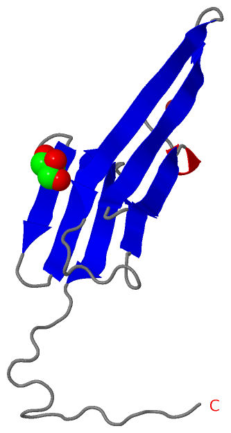
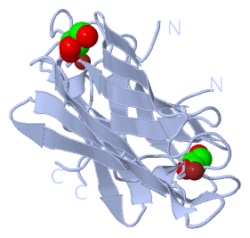
 Description
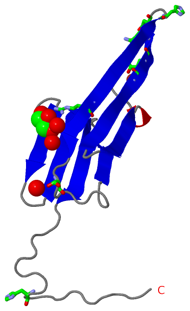
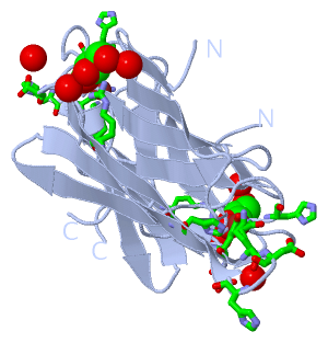