| molecular function |
|---|
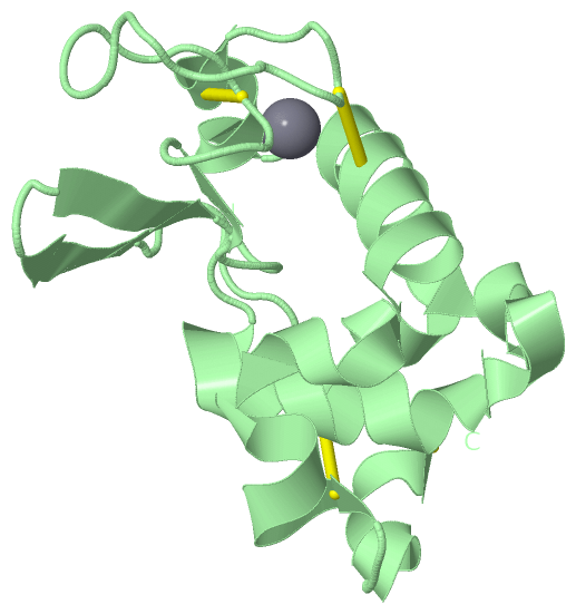
| | GO:0005509 | | calcium ion binding | | Interacting selectively and non-covalently with calcium ions (Ca2+). |
| | GO:0004461 | | lactose synthase activity | | Catalysis of the reaction: UDP-galactose + D-glucose = UDP + lactose. |
| | GO:0046872 | | metal ion binding | | Interacting selectively and non-covalently with any metal ion. |
| biological process |
|---|
| | GO:0006915 | | apoptotic process | | A programmed cell death process which begins when a cell receives an internal (e.g. DNA damage) or external signal (e.g. an extracellular death ligand), and proceeds through a series of biochemical events (signaling pathway phase) which trigger an execution phase. The execution phase is the last step of an apoptotic process, and is typically characterized by rounding-up of the cell, retraction of pseudopodes, reduction of cellular volume (pyknosis), chromatin condensation, nuclear fragmentation (karyorrhexis), plasma membrane blebbing and fragmentation of the cell into apoptotic bodies. When the execution phase is completed, the cell has died. |
| | GO:0007267 | | cell-cell signaling | | Any process that mediates the transfer of information from one cell to another. This process includes signal transduction in the receiving cell and, where applicable, release of a ligand and any processes that actively facilitate its transport and presentation to the receiving cell. Examples include signaling via soluble ligands, via cell adhesion molecules and via gap junctions. |
| | GO:0042742 | | defense response to bacterium | | Reactions triggered in response to the presence of a bacterium that act to protect the cell or organism. |
| | GO:0005989 | | lactose biosynthetic process | | The chemical reactions and pathways resulting in the formation of lactose, the disaccharide galactopyranosyl-glucose. |
| | GO:0007165 | | signal transduction | | The cellular process in which a signal is conveyed to trigger a change in the activity or state of a cell. Signal transduction begins with reception of a signal (e.g. a ligand binding to a receptor or receptor activation by a stimulus such as light), or for signal transduction in the absence of ligand, signal-withdrawal or the activity of a constitutively active receptor. Signal transduction ends with regulation of a downstream cellular process, e.g. regulation of transcription or regulation of a metabolic process. Signal transduction covers signaling from receptors located on the surface of the cell and signaling via molecules located within the cell. For signaling between cells, signal transduction is restricted to events at and within the receiving cell. |
| cellular component |
|---|
| | GO:0005796 | | Golgi lumen | | The volume enclosed by the membranes of any cisterna or subcompartment of the Golgi apparatus, including the cis- and trans-Golgi networks. |
| | GO:0000139 | | Golgi membrane | | The lipid bilayer surrounding any of the compartments of the Golgi apparatus. |
| | GO:0005576 | | extracellular region | | The space external to the outermost structure of a cell. For cells without external protective or external encapsulating structures this refers to space outside of the plasma membrane. This term covers the host cell environment outside an intracellular parasite. |
| | GO:0005615 | | extracellular space | | That part of a multicellular organism outside the cells proper, usually taken to be outside the plasma membranes, and occupied by fluid. |
 Description