Show PDB file:   
         Plain Text   HTML   (compressed file size)
QuickSearch:   
by PDB,NDB,UniProt,PROSITE Code or Search Term(s)  
(-)Asym.Unit - manually
(-)Asymmetric Unit
(-)Biological Unit 1
collapse expand < >
Image Asym.Unit - manually
Asym.Unit - manually  (Jmol Viewer)
Image Asymmetric Unit
Asymmetric Unit  (Jmol Viewer)
Image Biological Unit 1
Biological Unit 1  (Jmol Viewer)

(-) Description

Title :  CRYSTAL STRUCTURE OF CAULOBACTER CRESCENTUS SSPB ORTHOLOG
 
Authors :  P. Chien, R. A. Grant, R. T. Sauer, T. A. Baker
Date :  15 Jun 07  (Deposition) - 04 Sep 07  (Release) - 24 Feb 09  (Revision)
Method :  X-RAY DIFFRACTION
Resolution :  2.55
Chains :  Asym. Unit :  A,B
Biol. Unit 1:  A,B  (2x)
Keywords :  Sspb, Adaptor, Clpx, Ssra, Unknown Function, Hydrolase Activator (Keyword Search: [Gene Ontology, PubMed, Web (Google))
 
Reference :  P. Chien, R. A. Grant, R. T. Sauer, T. A. Baker
Crystal Structure Of Caulobacter Crescentus Sspb Ortholog
To Be Published
PubMed: search
(for further references see the PDB file header)

(-) Compounds

Molecule 1 - HYPOTHETICAL PROTEIN
    ChainsA
    EngineeredYES
    Expression SystemESCHERICHIA COLI BL21(DE3)
    Expression System PlasmidPET28
    Expression System StrainBL21(DE3)
    Expression System Taxid469008
    Expression System Vector TypePLASMID
    GeneCC_2102
    Organism ScientificCAULOBACTER VIBRIOIDES
    Organism Taxid155892
    StrainCB15N
    SynonymSSPB
 
Molecule 2 - C. CRESCENTUS SSRA PEPTIDE
    ChainsB
    EngineeredYES
    Other DetailsSYNTHETIC SSRA PEPITDE WITH N-TERMINAL SOLUBILIZATION TAG
    SyntheticYES

 Structural Features

(-) Chains, Units

  12
Asymmetric Unit AB
Biological Unit 1 (2x)AB

Summary Information (see also Sequences/Alignments below)

(-) Ligands, Modified Residues, Ions  (0, 0)

(no "Ligand,Modified Residues,Ions" information available for 2QAS)

(-) Sites  (0, 0)

(no "Site" information available for 2QAS)

(-) SS Bonds  (0, 0)

(no "SS Bond" information available for 2QAS)

(-) Cis Peptide Bonds  (2, 2)
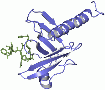
Asymmetric Unit
No.Residues
1Pro A:6 -Pro A:7
2Glu A:40 -Pro A:41

 Sequence-Structure Mapping

(-) SAPs(SNPs)/Variants  (0, 0)

(no "SAP(SNP)/Variant" information available for 2QAS)

(-) PROSITE Motifs  (0, 0)

(no "PROSITE Motif" information available for 2QAS)

(-) Exons   (0, 0)

(no "Exon" information available for 2QAS)

(-) Sequences/Alignments

Asymmetric Unit
   Reformat: Number of residues per line =  ('0' or empty: single-line sequence representation)
  Number of residues per labelling interval =   
  UniProt sequence: complete  aligned part    
   Show mapping: SCOP domains CATH domains Pfam domains Secondary structure (by author)
SAPs(SNPs) PROSITE motifs Exons
(details for a mapped element are shown in a popup box when the mouse pointer rests over it)
Chain A from PDB  Type:PROTEIN  Length:120
 aligned with Q9A6J2_CAUCR | Q9A6J2 from UniProtKB/TrEMBL  Length:162

    Alignment length:120
                                    15        25        35        45        55        65        75        85        95       105       115       125
         Q9A6J2_CAUCR     6 PPEDLMQYEAMAQDALRGVVKAALKKAAAPGGLPEPHHLYITFKTKAAGVSGPQDLLSKYPDEMTIVLQHQYWDLAPGETFFSVTLKFGGQPKRLSVPYAALTRFYDPSVQFALQFSAPE 125
               SCOP domains ------------------------------------------------------------------------------------------------------------------------ SCOP domains
               CATH domains 2qasA00 A:6-125 Stringent starvation protein B, SspB                                                                     CATH domains
               Pfam domains -----SspB-2qasA01 A:11-125                                                                                               Pfam domains
         Sec.struct. author ......hhhhhhhhhhhhhhhhhhhhhh..........eeeeee........hhhhhhhh..eeeeee....eeeee...eeeeeeee..eeeeeeee...eeeeee....eeee..... Sec.struct. author
                 SAPs(SNPs) ------------------------------------------------------------------------------------------------------------------------ SAPs(SNPs)
                    PROSITE ------------------------------------------------------------------------------------------------------------------------ PROSITE
                 Transcript ------------------------------------------------------------------------------------------------------------------------ Transcript
                 2qas A   6 PPEDLMQYEAMAQDALRGVVKAALKKAAAPGGLPEPHHLYITFKTKAAGVSGPQDLLSKYPDEMTIVLQHQYWDLAPGETFFSVTLKFGGQPKRLSVPYAALTRFYDPSVQFALQFSAPE 125
                                    15        25        35        45        55        65        75        85        95       105       115       125

Chain B from PDB  Type:PROTEIN  Length:10
                                          
               SCOP domains ---------- SCOP domains
               CATH domains ---------- CATH domains
               Pfam domains ---------- Pfam domains
         Sec.struct. author .......... Sec.struct. author
                 SAPs(SNPs) ---------- SAPs(SNPs)
                    PROSITE ---------- PROSITE
                 Transcript ---------- Transcript
                 2qas B   6 HGAANDNFAE  15
                                    15

   Legend:   → Mismatch (orange background)
  - → Gap (green background, '-', border residues have a numbering label)
    → Modified Residue (blue background, lower-case, 'x' indicates undefined single-letter code, labelled with number + name)
  x → Chemical Group (purple background, 'x', labelled with number + name, e.g. ACE or NH2)
  extra numbering lines below/above indicate numbering irregularities and modified residue names etc., number ends below/above '|'

 Classification and Annotation

(-) SCOP Domains  (0, 0)

(no "SCOP Domain" information available for 2QAS)

(-) CATH Domains  (1, 1)

Asymmetric Unit
(-)
Class: Mainly Beta (13760)

(-) Pfam Domains  (1, 1)

Asymmetric Unit

(-) Gene Ontology  (0, 0)

Asymmetric Unit(hide GO term definitions)
    (no "Gene Ontology" information available for 2QAS)

 Visualization

(-) Interactive Views

Asymmetric Unit
  Complete Structure
    Jena3D(integrated viewing of ligand, site, SAP, PROSITE, SCOP information)
    WebMol | AstexViewer[tm]@PDBe
(Java Applets, require no local installation except for Java; loading may be slow)
    STRAP
(Java WebStart application, automatic local installation, requires Java; full application with system access!)
    RasMol
(require local installation)
    Molscript (VRML)
(requires installation of a VRML viewer; select preferred view via VRML and generate a mono or stereo PDF format file)
 
  Ligands, Modified Residues, Ions
(no "Ligands, Modified Residues, Ions" information available for 2qas)
 
  Sites
(no "Sites" information available for 2qas)
 
  Cis Peptide Bonds
    Glu A:40 - Pro A:41   [ RasMol ]  
    Pro A:6 - Pro A:7   [ RasMol ]  
 
Biological Unit
  Complete Structure
    Biological Unit 1  [ Jena3D ]

(-) Still Images

Jmol
  protein: cartoon or spacefill or dots and stick; nucleic acid: cartoon and stick; ligands: spacefill; active site: stick
Molscript
  protein, nucleic acid: cartoon; ligands: spacefill; active site: ball and stick

 Databases and Analysis Tools

(-) Databases

Access by PDB/NDB ID
  2qas
    Family and Domain InformationProDom | SYSTERS
    General Structural InformationGlycoscienceDB | MMDB | NDB | OCA | PDB | PDBe | PDBj | PDBsum | PDBWiki | PQS | PROTEOPEDIA
    Orientation in MembranesOPM
    Protein SurfaceSURFACE
    Secondary StructureDSSP (structure derived) | HSSP (homology derived)
    Structural GenomicsGeneCensus
    Structural NeighboursCE | VAST
    Structure ClassificationCATH | Dali | SCOP
    Validation and Original DataBMRB Data View | BMRB Restraints Grid | EDS | PROCHECK | RECOORD | WHAT_CHECK
 
Access by UniProt ID/Accession number
  Q9A6J2_CAUCR | Q9A6J2
    Comparative Protein Structure ModelsModBase
    Genomic InformationEnsembl
    Protein-protein InteractionDIP
    Sequence, Family and Domain InformationInterPro | Pfam | SMART | UniProtKB/TrEMBL
 
Access by Enzyme Classificator   (EC Number)
  (no 'Enzyme Classificator' available)
    General Enzyme InformationBRENDA | EC-PDB | Enzyme | IntEnz
    PathwayKEGG | MetaCyc
 
Access by Disease Identifier   (MIM ID)
  (no 'MIM ID' available)
    Disease InformationOMIM
 
Access by GenAge ID
  (no 'GenAge ID' available)
    Age Related InformationGenAge

(-) Analysis Tools

Access by PDB/NDB ID
    Domain InformationXDom
    Interatomic Contacts of Structural UnitsCSU
    Ligand-protein ContactsLPC
    Protein CavitiescastP
    Sequence and Secondary StructurePDBCartoon
    Structure AlignmentSTRAP(Java WebStart application, automatic local installation, requires Java; full application with system access!)
    Structure and Sequence BrowserSTING
 
Access by UniProt ID/Accession number
  Q9A6J2_CAUCR | Q9A6J2
    Protein Disorder PredictionDisEMBL | FoldIndex | GLOBPLOT (for more information see DisProt)

 Related Entries

(-) Entries Sharing at Least One Protein Chain (UniProt ID)

UniProtKB/TrEMBL
        Q9A6J2_CAUCR | Q9A6J22qaz

(-) Related Entries Specified in the PDB File

2nys ORTHOLOGOUS PROTEIN FROM AGROBACTERIA
2qaz