Show PDB file:   
         Plain Text   HTML   (compressed file size)
QuickSearch:   
by PDB,NDB,UniProt,PROSITE Code or Search Term(s)  
(-)Asym.Unit - manually
(-)Asymmetric Unit
(-)Asym. Unit - sites
(-)Biological Unit 1
(-)Biol. Unit 1 - sites
(-)Biological Unit 2
collapse expand < >
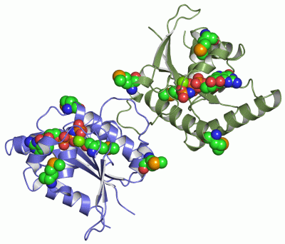
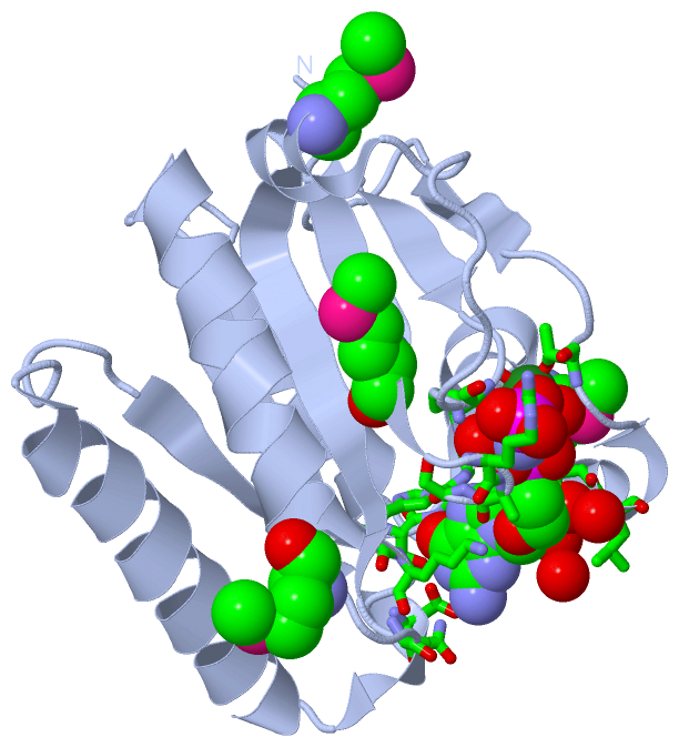
Image Asym.Unit - manually
Asym.Unit - manually  (Jmol Viewer)
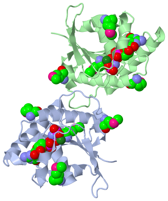
Image Asymmetric Unit
Asymmetric Unit  (Jmol Viewer)
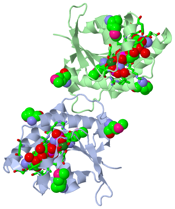
Image Asym. Unit - sites
Asym. Unit - sites  (Jmol Viewer)
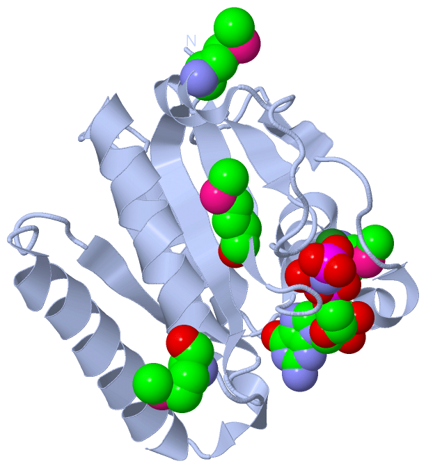
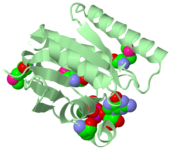
Image Biological Unit 1
Biological Unit 1  (Jmol Viewer)
Image Biol. Unit 1 - sites
Biol. Unit 1 - sites  (Jmol Viewer)
Image Biological Unit 2
Biological Unit 2  (Jmol Viewer)

(-) Description

Title :  X-RAY CRYSTAL STRUCTURE OF PUTATIVE HUMAN RAS-RELATED GTP BINDING D IN COMPLEX WITH GMPPNP
 
Authors :  A. M. Mulichak, W. M. Rabeh, W. Tempel, L. Nedyalkova, R. Landry, C. H. Arrowsmith, A. M. Edwards, M. Sundstrom, J. Weigelt, L. J. Keefe, A. Bochkarev, H. Park, Structural Genomics Consortium (Sgc)
Date :  30 May 07  (Deposition) - 12 Jun 07  (Release) - 24 Feb 09  (Revision)
Method :  X-RAY DIFFRACTION
Resolution :  2.10
Chains :  Asym. Unit :  A,B
Biol. Unit 1:  A  (1x)
Biol. Unit 2:  B  (1x)
Keywords :  Structural Genomics, Gtp-Binding, Rragd, Structural Genomics Consortium, Sgc, Protein Binding (Keyword Search: [Gene Ontology, PubMed, Web (Google)] )
 
Reference :  A. M. Mulichak, W. M. Rabeh, W. Tempel, L. Nedyalkova, R. Landry, C. H. Arrowsmith, A. M. Edwards, M. Sundstrom, J. Weigelt, L. J. Keefe, A. Bochkarev, H. Park
Crystal Structure Of Human Ras-Related Gtp-Binding D.
To Be Published
PubMed: search
(for further references see the PDB file header)

(-) Compounds

Molecule 1 - RAS-RELATED GTP-BINDING PROTEIN D
    Chains: A, B
    Engineered: YES
    Expression System: ESCHERICHIA COLI BL21(DE3)
    Expression System Plasmid: P28A-LIC
    Expression System Strain: BL21 (DE3)
    Expression System Taxid: 469008
    Expression System Vector Type: PLASMID
    Gene: RRAGD
    Organism Common: HUMAN
    Organism Scientific: HOMO SAPIENS
    Organism Taxid: 9606
    Synonym: RAG D

 Structural Features

(-) Chains, Units

  12
Asymmetric Unit : AB
Biological Unit 1 (1x): A 
Biological Unit 2 (1x):  B

Summary Information (see also Sequences/Alignments below)

(-) Ligands, Modified Residues, Ions  (3, 12)

Asymmetric Unit (3, 12)
No.NameCountTypeFull Name
1GNP2Ligand/IonPHOSPHOAMINOPHOSPHONIC ACID-GUANYLATE ESTER
2MG2Ligand/IonMAGNESIUM ION
3MSE8Mod. Amino AcidSELENOMETHIONINE
Biological Unit 1 (2, 5)
No.NameCountTypeFull Name
1GNP1Ligand/IonPHOSPHOAMINOPHOSPHONIC ACID-GUANYLATE ESTER
2MG-1Ligand/IonMAGNESIUM ION
3MSE4Mod. Amino AcidSELENOMETHIONINE
Biological Unit 2 (2, 5)
No.NameCountTypeFull Name
1GNP1Ligand/IonPHOSPHOAMINOPHOSPHONIC ACID-GUANYLATE ESTER
2MG-1Ligand/IonMAGNESIUM ION
3MSE4Mod. Amino AcidSELENOMETHIONINE

(-) Sites  (4, 4)

Asymmetric Unit (4, 4)
No.NameEvidenceResiduesDescription
1AC1SOFTWARESER A:57 , THR A:78 , GNP A:401 , HOH A:406 , HOH A:408BINDING SITE FOR RESIDUE MG A 301
2AC2SOFTWARESER B:57 , THR B:78 , GNP B:402 , HOH B:415 , HOH B:425BINDING SITE FOR RESIDUE MG B 302
3AC3SOFTWAREARG A:52 , ARG A:53 , SER A:54 , GLY A:55 , LYS A:56 , SER A:57 , SER A:58 , THR A:72 , LEU A:73 , THR A:78 , GLY A:101 , GLN A:129 , HIS A:160 , LYS A:161 , ASP A:163 , SER A:201 , ILE A:202 , MG A:301 , HOH A:406 , HOH A:408 , HOH A:422 , HOH A:430BINDING SITE FOR RESIDUE GNP A 401
4AC4SOFTWARELEU B:51 , ARG B:52 , ARG B:53 , SER B:54 , GLY B:55 , LYS B:56 , SER B:57 , SER B:58 , THR B:72 , LEU B:73 , THR B:78 , GLY B:101 , HIS B:160 , LYS B:161 , ASP B:163 , SER B:201 , ILE B:202 , TYR B:203 , MG B:302 , HOH B:410 , HOH B:415 , HOH B:417 , HOH B:425 , HOH B:440BINDING SITE FOR RESIDUE GNP B 402

(-) SS Bonds  (0, 0)

(no "SS Bond" information available for 2Q3F)

(-) Cis Peptide Bonds  (0, 0)

(no "Cis Peptide Bond" information available for 2Q3F)

 Sequence-Structure Mapping

(-) SAPs(SNPs)/Variants  (0, 0)

(no "SAP(SNP)/Variant" information available for 2Q3F)

(-) PROSITE Motifs  (0, 0)

(no "PROSITE Motif" information available for 2Q3F)

(-) Exons   (3, 6)

Asymmetric Unit (3, 6)
 ENSEMBLUniProtKBPDB
No.Transcript IDExonExon IDGenome LocationLengthIDLocationLengthCountLocationLength
1.1aENST000003694151aENSE00001449989chr6:90121989-90121565425RRAGD_HUMAN1-50500--
1.3ENST000003694153ENSE00002197569chr6:90097309-90097014296RRAGD_HUMAN50-148992A:42-129
B:42-129
88
88
1.4aENST000003694154aENSE00002195650chr6:90090086-90089887200RRAGD_HUMAN149-215672A:130-196
B:130-196
67
67
1.5ENST000003694155ENSE00002151390chr6:90089057-90088943115RRAGD_HUMAN215-253392A:196-220
B:196-219
25
24
1.6bENST000003694156bENSE00001888083chr6:90087532-90087390143RRAGD_HUMAN254-301480--
1.7ENST000003694157ENSE00000760434chr6:90082304-90082156149RRAGD_HUMAN301-351510--
1.8bENST000003694158bENSE00001449984chr6:90077926-900743553572RRAGD_HUMAN351-400500--

(-) Sequences/Alignments

Asymmetric Unit
   Reformat: Number of residues per line =  ('0' or empty: single-line sequence representation)
  Number of residues per labelling interval =   
  UniProt sequence: complete  aligned part    
   Show mapping: SCOP domains CATH domains Pfam domains Secondary structure (by author)
SAPs(SNPs) PROSITE motifs Exons
(details for a mapped element are shown in a popup box when the mouse pointer rests over it)
Chain A from PDB  Type:PROTEIN  Length:179
 aligned with RRAGD_HUMAN | Q9NQL2 from UniProtKB/Swiss-Prot  Length:400

    Alignment length:179
                                    70        80        90       100       110       120       130       140       150       160       170       180       190       200       210       220       230         
          RRAGD_HUMAN    61 VKPRILLMGLRRSGKSSIQKVVFHKMSPNETLFLESTNKICREDVSNSSFVNFQIWDFPGQIDFFDPTFDYEMIFRGTGALIFVIDSQDDYMEALARLHLTVTRAYKVNTDINFEVFIHKVDGLSDDHKIETQRDIHQRANDDLADAGLEKIHLSFYLTSIYDHSIFEAFSKVVQKLIP 239
               SCOP domains ----------------------------------------------------------------------------------------------------------------------------------------------------------------------------------- SCOP domains
               CATH domains ----------------------------------------------------------------------------------------------------------------------------------------------------------------------------------- CATH domains
               Pfam domains ----------------------------------------------------------------------------------------------------------------------------------------------------------------------------------- Pfam domains
         Sec.struct. author ...eeeeee....hhhhhhhhhhhh.hhhhhhhh......eeeee.......eeeee............hhhhhhhheeeeeeeee....hhhhhhhhhhhhhhhhhhh...eeeeeee.hhhh..hhhhhhhhhhhhhhhhhhhhh......eeeeee....hhhhhhhhhhhhh... Sec.struct. author
                 SAPs(SNPs) ----------------------------------------------------------------------------------------------------------------------------------------------------------------------------------- SAPs(SNPs)
                    PROSITE ----------------------------------------------------------------------------------------------------------------------------------------------------------------------------------- PROSITE
           Transcript 1 (1) Exon 1.3  PDB: A:42-129 UniProt: 50-148 [INCOMPLETE]                                    Exon 1.4a  PDB: A:130-196 UniProt: 149-215                         ------------------------ Transcript 1 (1)
           Transcript 1 (2) ----------------------------------------------------------------------------------------------------------------------------------------------------------Exon 1.5  PDB: A:196-220  Transcript 1 (2)
                 2q3f A  42 VKPRILLmGLRRSGKSSIQKVVFHKmSPNETLFLESTNKICREDVSNSSFVNFQIWDFPGQIDFFDPTFDYEmIFRGTGALIFVIDSQDDYmEALARLHLTVTRAYKVNTDINFEVFIHKVDGLSDDHKIETQRDIHQRANDDLADAGLEKIHLSFYLTSIYDHSIFEAFSKVVQKLIP 220
                                   |51        61     |  71        81        91       101       111  |    121       131 |     141       151       161       171       181       191       201       211         
                                  49-MSE            67-MSE                                        114-MSE            133-MSE                                                                                   

Chain B from PDB  Type:PROTEIN  Length:178
 aligned with RRAGD_HUMAN | Q9NQL2 from UniProtKB/Swiss-Prot  Length:400

    Alignment length:178
                                    70        80        90       100       110       120       130       140       150       160       170       180       190       200       210       220       230        
          RRAGD_HUMAN    61 VKPRILLMGLRRSGKSSIQKVVFHKMSPNETLFLESTNKICREDVSNSSFVNFQIWDFPGQIDFFDPTFDYEMIFRGTGALIFVIDSQDDYMEALARLHLTVTRAYKVNTDINFEVFIHKVDGLSDDHKIETQRDIHQRANDDLADAGLEKIHLSFYLTSIYDHSIFEAFSKVVQKLI 238
               SCOP domains ---------------------------------------------------------------------------------------------------------------------------------------------------------------------------------- SCOP domains
               CATH domains ---------------------------------------------------------------------------------------------------------------------------------------------------------------------------------- CATH domains
           Pfam domains (1) ---Gtr1_RagA-2q3fB01 B:45-219                                                                                                                                                      Pfam domains (1)
           Pfam domains (2) ---Gtr1_RagA-2q3fB02 B:45-219                                                                                                                                                      Pfam domains (2)
         Sec.struct. author ...eeeee.....hhhhhhhhhhh..hhhhhhhh......eeeee.......eeeee............hhhhhhhheeeeeeeee....hhhhhhhhhhhhhhhhhhh...eeeeeee.....hhhhhhhhhhhhhhhhhhhhhhh......eeeee.....hhhhhhhhhhhhh.. Sec.struct. author
                 SAPs(SNPs) ---------------------------------------------------------------------------------------------------------------------------------------------------------------------------------- SAPs(SNPs)
                    PROSITE ---------------------------------------------------------------------------------------------------------------------------------------------------------------------------------- PROSITE
           Transcript 1 (1) Exon 1.3  PDB: B:42-129 UniProt: 50-148 [INCOMPLETE]                                    Exon 1.4a  PDB: B:130-196 UniProt: 149-215                         ----------------------- Transcript 1 (1)
           Transcript 1 (2) ----------------------------------------------------------------------------------------------------------------------------------------------------------Exon 1.5  PDB: B:196-219 Transcript 1 (2)
                 2q3f B  42 VKPRILLmGLRRSGKSSIQKVVFHKmSPNETLFLESTNKICREDVSNSSFVNFQIWDFPGQIDFFDPTFDYEmIFRGTGALIFVIDSQDDYmEALARLHLTVTRAYKVNTDINFEVFIHKVDGLSDDHKIETQRDIHQRANDDLADAGLEKIHLSFYLTSIYDHSIFEAFSKVVQKLI 219
                                   |51        61     |  71        81        91       101       111  |    121       131 |     141       151       161       171       181       191       201       211        
                                  49-MSE            67-MSE                                        114-MSE            133-MSE                                                                                  

   Legend:   → Mismatch (orange background)
  - → Gap (green background, '-', border residues have a numbering label)
    → Modified Residue (blue background, lower-case, 'x' indicates undefined single-letter code, labelled with number + name)
  x → Chemical Group (purple background, 'x', labelled with number + name, e.g. ACE or NH2)
  extra numbering lines below/above indicate numbering irregularities and modified residue names etc., number ends below/above '|'

 Classification and Annotation

(-) SCOP Domains  (0, 0)

(no "SCOP Domain" information available for 2Q3F)

(-) CATH Domains  (0, 0)

(no "CATH Domain" information available for 2Q3F)

(-) Pfam Domains  (1, 2)

Asymmetric Unit

(-) Gene Ontology  (18, 18)

Asymmetric Unit(hide GO term definitions)
Chain A,B   (RRAGD_HUMAN | Q9NQL2)
molecular function
    GO:0005525    GTP binding    Interacting selectively and non-covalently with GTP, guanosine triphosphate.
    GO:0003924    GTPase activity    Catalysis of the reaction: GTP + H2O = GDP + phosphate.
    GO:0000166    nucleotide binding    Interacting selectively and non-covalently with a nucleotide, any compound consisting of a nucleoside that is esterified with (ortho)phosphate or an oligophosphate at any hydroxyl group on the ribose or deoxyribose.
    GO:0005515    protein binding    Interacting selectively and non-covalently with any protein or protein complex (a complex of two or more proteins that may include other nonprotein molecules).
    GO:0046982    protein heterodimerization activity    Interacting selectively and non-covalently with a nonidentical protein to form a heterodimer.
biological process
    GO:0007050    cell cycle arrest    A regulatory process that halts progression through the cell cycle during one of the normal phases (G1, S, G2, M).
    GO:0034613    cellular protein localization    Any process in which a protein is transported to, and/or maintained in, a specific location at the level of a cell. Localization at the cellular level encompasses movement within the cell, from within the cell to the cell surface, or from one location to another at the surface of a cell.
    GO:0071230    cellular response to amino acid stimulus    Any process that results in a change in state or activity of a cell (in terms of movement, secretion, enzyme production, gene expression, etc.) as a result of an amino acid stimulus. An amino acid is a carboxylic acids containing one or more amino groups.
    GO:0009267    cellular response to starvation    Any process that results in a change in state or activity of a cell (in terms of movement, secretion, enzyme production, gene expression, etc.) as a result of deprivation of nourishment.
    GO:0016236    macroautophagy    The major inducible pathway for the general turnover of cytoplasmic constituents in eukaryotic cells, it is also responsible for the degradation of active cytoplasmic enzymes and organelles during nutrient starvation. Macroautophagy involves the formation of double-membrane-bounded autophagosomes which enclose the cytoplasmic constituent targeted for degradation in a membrane-bounded structure. Autophagosomes then fuse with a lysosome (or vacuole) releasing single-membrane-bounded autophagic bodies that are then degraded within the lysosome (or vacuole). Though once thought to be a purely non-selective process, it appears that some types of macroautophagy, e.g. macropexophagy, macromitophagy, may involve selective targeting of the targets to be degraded.
    GO:0032008    positive regulation of TOR signaling    Any process that activates or increases the frequency, rate or extent of TOR signaling.
    GO:0010506    regulation of autophagy    Any process that modulates the frequency, rate or extent of autophagy. Autophagy is the process in which cells digest parts of their own cytoplasm.
cellular component
    GO:0034448    EGO complex    A vacuolar membrane-associated protein complex that is required for activation of microautophagy during exit from rapamycin-induced growth arrest. In budding yeast, S. cerevisiae, this complex includes Gtr1p, Gtr2p, Meh1p, and Slm4p.
    GO:1990131    Gtr1-Gtr2 GTPase complex    A heterodimer GTPase complex. In S. cerevisiae, this complex contains Gtr1p and Gtr2p proteins.
    GO:0005737    cytoplasm    All of the contents of a cell excluding the plasma membrane and nucleus, but including other subcellular structures.
    GO:0005829    cytosol    The part of the cytoplasm that does not contain organelles but which does contain other particulate matter, such as protein complexes.
    GO:0005764    lysosome    A small lytic vacuole that has cell cycle-independent morphology and is found in most animal cells and that contains a variety of hydrolases, most of which have their maximal activities in the pH range 5-6. The contained enzymes display latency if properly isolated. About 40 different lysosomal hydrolases are known and lysosomes have a great variety of morphologies and functions.
    GO:0005634    nucleus    A membrane-bounded organelle of eukaryotic cells in which chromosomes are housed and replicated. In most cells, the nucleus contains all of the cell's chromosomes except the organellar chromosomes, and is the site of RNA synthesis and processing. In some species, or in specialized cell types, RNA metabolism or DNA replication may be absent.

 Visualization

(-) Interactive Views

Asymmetric Unit
  Complete Structure
    Jena3D(integrated viewing of ligand, site, SAP, PROSITE, SCOP information)
    WebMol | AstexViewer[tm]@PDBe
(Java Applets, require no local installation except for Java; loading may be slow)
    STRAP
(Java WebStart application, automatic local installation, requires Java; full application with system access!)
    RasMol
(require local installation)
    Molscript (VRML)
(requires installation of a VRML viewer; select preferred view via VRML and generate a mono or stereo PDF format file)
 
  Ligands, Modified Residues, Ions
    GNP  [ RasMol | Jena3D ]  +environment [ RasMol | Jena3D ]
    MG  [ RasMol | Jena3D ]  +environment [ RasMol | Jena3D ]
    MSE  [ RasMol | Jena3D ]  +environment [ RasMol | Jena3D ]
 
  Sites
    AC1  [ RasMol ]  +environment [ RasMol ]
    AC2  [ RasMol ]  +environment [ RasMol ]
    AC3  [ RasMol ]  +environment [ RasMol ]
    AC4  [ RasMol ]  +environment [ RasMol ]
 
  Cis Peptide Bonds
(no "Cis Peptide Bonds" information available for 2q3f)
 
Biological Units
  Complete Structure
    Biological Unit 1  [ Jena3D ]
    Biological Unit 2  [ Jena3D ]

(-) Still Images

Jmol
  protein: cartoon or spacefill or dots and stick; nucleic acid: cartoon and stick; ligands: spacefill; active site: stick
Molscript
  protein, nucleic acid: cartoon; ligands: spacefill; active site: ball and stick

 Databases and Analysis Tools

(-) Databases

Access by PDB/NDB ID
  2q3f
    Family and Domain Information: ProDom | SYSTERS
    General Structural Information: GlycoscienceDB | MMDB | NDB | OCA | PDB | PDBe | PDBj | PDBsum | PDBWiki | PQS | PROTEOPEDIA
    Orientation in Membranes: OPM
    Protein Surface: SURFACE
    Secondary Structure: DSSP (structure derived) | HSSP (homology derived)
    Structural Genomics: GeneCensus
    Structural Neighbours: CE | VAST
    Structure Classification: CATH | Dali | SCOP
    Validation and Original Data: BMRB Data View | BMRB Restraints Grid | EDS | PROCHECK | RECOORD | WHAT_CHECK
 
Access by UniProt ID/Accession number
  RRAGD_HUMAN | Q9NQL2
    Comparative Protein Structure Models: ModBase
    Genomic Information: Ensembl
    Protein-protein Interaction: DIP
    Sequence, Family and Domain Information: InterPro | Pfam | SMART | UniProtKB/SwissProt
 
Access by Enzyme Classificator   (EC Number)
  (no 'Enzyme Classificator' available)
    General Enzyme Information: BRENDA | EC-PDB | Enzyme | IntEnz
    Pathway: KEGG | MetaCyc
 
Access by Disease Identifier   (MIM ID)
  (no 'MIM ID' available)
    Disease Information: OMIM
 
Access by GenAge ID
  (no 'GenAge ID' available)
    Age Related Information: GenAge

(-) Analysis Tools

Access by PDB/NDB ID
    Domain Information: XDom
    Interatomic Contacts of Structural Units: CSU
    Ligand-protein Contacts: LPC
    Protein Cavities: castP
    Sequence and Secondary Structure: PDBCartoon
    Structure Alignment: STRAP(Java WebStart application, automatic local installation, requires Java; full application with system access!)
    Structure and Sequence Browser: STING
 
Access by UniProt ID/Accession number
  RRAGD_HUMAN | Q9NQL2
    Protein Disorder Prediction: DisEMBL | FoldIndex | GLOBPLOT (for more information see DisProt)

 Related Entries

(-) Entries Sharing at Least One Protein Chain (UniProt ID)

(no "Entries Sharing at Least One Protein Chain" available for 2Q3F)

(-) Related Entries Specified in the PDB File

(no "Related Entries Specified in the PDB File" available for 2Q3F)