Show PDB file:   
         Plain Text   HTML   (compressed file size)
QuickSearch:   
by PDB,NDB,UniProt,PROSITE Code or Search Term(s)  
(-)Asym./Biol. Unit
collapse expand < >
Image Asym./Biol. Unit
Asym./Biol. Unit  (Jmol Viewer)

(-) Description

Title :  CRYSTAL STRUCTURE OF THE Q4KFT4_PSEF5 PROTEIN FROM PSEUDOMONAS FLUORESCENS. NESG TARGET PLR1
 
Authors :  S. M. Vorobiev, H. Neely, S. Jayaraman, C. X. Chen, H. Janjua, R. Xiao, T. G. T. Montelione, J. F. Hunt, L. Tong, Northeast Structural Genomic Consortium (Nesg)
Date :  17 Apr 07  (Deposition) - 01 May 07  (Release) - 13 Jul 11  (Revision)
Method :  X-RAY DIFFRACTION
Resolution :  2.20
Chains :  Asym./Biol. Unit :  A,B
Keywords :  Nesg, Plr1, Putative Tetraacyldisaccharide-1-P 4-Kinase, Q4Kft4, Structural Genomics, Psi-2, Protein Structure Initiative, Northeast Structural Genomics Consortium, Unknown Function (Keyword Search: [Gene Ontology, PubMed, Web (Google))
 
Reference :  S. M. Vorobiev, H. Neely, S. Jayaraman, C. X. Chen, H. Janjua, R. Xiao, T. Acton, G. T. Montelione, J. F. Hunt, L. Tong
Crystal Structure Of The Q4Kft4_psef5 Protein From Pseudomonas Fluorescens.
To Be Published
PubMed: search

(-) Compounds

Molecule 1 - UNCHARACTERIZED PROTEIN
    AtccBAA-477
    ChainsA, B
    EngineeredYES
    Expression SystemESCHERICHIA COLI
    Expression System PlasmidPET21
    Expression System StrainBL21(DE3)+MAGIC
    Expression System Taxid562
    Expression System Vector TypePLASMID
    GenePFL_1779
    Organism ScientificPSEUDOMONAS FLUORESCENS
    Organism Taxid220664
    StrainPF-5

 Structural Features

(-) Chains, Units

  12
Asymmetric/Biological Unit AB

Summary Information (see also Sequences/Alignments below)

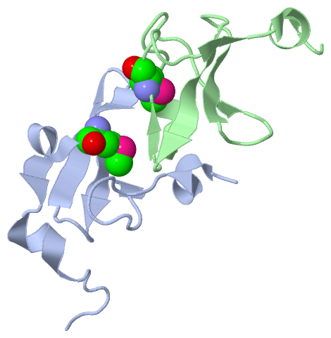
(-) Ligands, Modified Residues, Ions  (1, 2)

Asymmetric/Biological Unit (1, 2)
No.NameCountTypeFull Name
1MSE2Mod. Amino AcidSELENOMETHIONINE

(-) Sites  (0, 0)

(no "Site" information available for 2PK7)

(-) SS Bonds  (0, 0)

(no "SS Bond" information available for 2PK7)

(-) Cis Peptide Bonds  (0, 0)

(no "Cis Peptide Bond" information available for 2PK7)

 Sequence-Structure Mapping

(-) SAPs(SNPs)/Variants  (0, 0)

(no "SAP(SNP)/Variant" information available for 2PK7)

(-) PROSITE Motifs  (0, 0)

(no "PROSITE Motif" information available for 2PK7)

(-) Exons   (0, 0)

(no "Exon" information available for 2PK7)

(-) Sequences/Alignments

Asymmetric/Biological Unit
   Reformat: Number of residues per line =  ('0' or empty: single-line sequence representation)
  Number of residues per labelling interval =   
  UniProt sequence: complete  aligned part    
   Show mapping: SCOP domains CATH domains Pfam domains Secondary structure (by author)
SAPs(SNPs) PROSITE motifs Exons
(details for a mapped element are shown in a popup box when the mouse pointer rests over it)
Chain A from PDB  Type:PROTEIN  Length:64
 aligned with Y1779_PSEF5 | Q4KFT4 from UniProtKB/Swiss-Prot  Length:61

    Alignment length:64
                                                                                     61     
                                    12        22        32        42        52        |-    
           Y1779_PSEF5    3 TKLLDILACPICKGPLKLSADKTELISKGAGLAYPIRDGIPVMLESEARTLTTEERLDK-----  -
               SCOP domains d2pk7a1 A:3-61 Uncharacterized protein PFL1779             ----- SCOP domains
               CATH domains -----2pk7A01 A:8-66  [code=2.20.25.10, no name defined]          CATH domains
               Pfam domains ---------------------------------------------------------------- Pfam domains
         Sec.struct. author .hhhhh..........ee.....eeee....eeeeee..ee..hhhhhee.hhhhhh....... Sec.struct. author
                 SAPs(SNPs) ---------------------------------------------------------------- SAPs(SNPs)
                    PROSITE ---------------------------------------------------------------- PROSITE
                 Transcript ---------------------------------------------------------------- Transcript
                  2pk7 A  3 TKLLDILACPICKGPLKLSADKTELISKGAGLAYPIRDGIPVmLESEARTLTTEERLDKLEHHH 66
                                    12        22        32        42  |     52        62    
                                                                     45-MSE                 

Chain B from PDB  Type:PROTEIN  Length:60
 aligned with Y1779_PSEF5 | Q4KFT4 from UniProtKB/Swiss-Prot  Length:61

    Alignment length:60
                                                                                     61 
                                    12        22        32        42        52        |-
           Y1779_PSEF5    3 TKLLDILACPICKGPLKLSADKTELISKGAGLAYPIRDGIPVMLESEARTLTTEERLDK-  -
               SCOP domains d2pk7b_ B: Uncharacterized protein PFL1779                   SCOP domains
               CATH domains -----2pk7B01 B:8-62  [code=2.20.25.10, no name defined]      CATH domains
           Pfam domains (1) Trm112p-2pk7B01 B:3-41                 --------------------- Pfam domains (1)
           Pfam domains (2) Trm112p-2pk7B02 B:3-41                 --------------------- Pfam domains (2)
         Sec.struct. author .hhhhhh.........ee.....eeee....eeeeee..ee.......ee.hhhhhh... Sec.struct. author
                 SAPs(SNPs) ------------------------------------------------------------ SAPs(SNPs)
                    PROSITE ------------------------------------------------------------ PROSITE
                 Transcript ------------------------------------------------------------ Transcript
                  2pk7 B  3 TKLLDILACPICKGPLKLSADKTELISKGAGLAYPIRDGIPVmLESEARTLTTEERLDKL 62
                                    12        22        32        42  |     52        62
                                                                     45-MSE             

   Legend:   → Mismatch (orange background)
  - → Gap (green background, '-', border residues have a numbering label)
    → Modified Residue (blue background, lower-case, 'x' indicates undefined single-letter code, labelled with number + name)
  x → Chemical Group (purple background, 'x', labelled with number + name, e.g. ACE or NH2)
  extra numbering lines below/above indicate numbering irregularities and modified residue names etc., number ends below/above '|'

 Classification and Annotation

(-) SCOP Domains  (1, 2)

Asymmetric/Biological Unit

(-) CATH Domains  (1, 2)

Asymmetric/Biological Unit
(-)
Class: Mainly Beta (13760)

(-) Pfam Domains  (1, 2)

Asymmetric/Biological Unit

(-) Gene Ontology  (0, 0)

Asymmetric/Biological Unit(hide GO term definitions)
    (no "Gene Ontology" information available for 2PK7)

 Visualization

(-) Interactive Views

Asymmetric/Biological Unit
  Complete Structure
    Jena3D(integrated viewing of ligand, site, SAP, PROSITE, SCOP information)
    WebMol | AstexViewer[tm]@PDBe
(Java Applets, require no local installation except for Java; loading may be slow)
    STRAP
(Java WebStart application, automatic local installation, requires Java; full application with system access!)
    RasMol
(require local installation)
    Molscript (VRML)
(requires installation of a VRML viewer; select preferred view via VRML and generate a mono or stereo PDF format file)
 
  Ligands, Modified Residues, Ions
    MSE  [ RasMol | Jena3D ]  +environment [ RasMol | Jena3D ]
 
  Sites
(no "Sites" information available for 2pk7)
 
  Cis Peptide Bonds
(no "Cis Peptide Bonds" information available for 2pk7)
 

(-) Still Images

Jmol
  protein: cartoon or spacefill or dots and stick; nucleic acid: cartoon and stick; ligands: spacefill; active site: stick
Molscript
  protein, nucleic acid: cartoon; ligands: spacefill; active site: ball and stick

 Databases and Analysis Tools

(-) Databases

Access by PDB/NDB ID
  2pk7
    Family and Domain InformationProDom | SYSTERS
    General Structural InformationGlycoscienceDB | MMDB | NDB | OCA | PDB | PDBe | PDBj | PDBsum | PDBWiki | PQS | PROTEOPEDIA
    Orientation in MembranesOPM
    Protein SurfaceSURFACE
    Secondary StructureDSSP (structure derived) | HSSP (homology derived)
    Structural GenomicsGeneCensus
    Structural NeighboursCE | VAST
    Structure ClassificationCATH | Dali | SCOP
    Validation and Original DataBMRB Data View | BMRB Restraints Grid | EDS | PROCHECK | RECOORD | WHAT_CHECK
 
Access by UniProt ID/Accession number
  Y1779_PSEF5 | Q4KFT4
    Comparative Protein Structure ModelsModBase
    Genomic InformationEnsembl
    Protein-protein InteractionDIP
    Sequence, Family and Domain InformationInterPro | Pfam | SMART | UniProtKB/SwissProt
 
Access by Enzyme Classificator   (EC Number)
  (no 'Enzyme Classificator' available)
    General Enzyme InformationBRENDA | EC-PDB | Enzyme | IntEnz
    PathwayKEGG | MetaCyc
 
Access by Disease Identifier   (MIM ID)
  (no 'MIM ID' available)
    Disease InformationOMIM
 
Access by GenAge ID
  (no 'GenAge ID' available)
    Age Related InformationGenAge

(-) Analysis Tools

Access by PDB/NDB ID
    Domain InformationXDom
    Interatomic Contacts of Structural UnitsCSU
    Ligand-protein ContactsLPC
    Protein CavitiescastP
    Sequence and Secondary StructurePDBCartoon
    Structure AlignmentSTRAP(Java WebStart application, automatic local installation, requires Java; full application with system access!)
    Structure and Sequence BrowserSTING
 
Access by UniProt ID/Accession number
  Y1779_PSEF5 | Q4KFT4
    Protein Disorder PredictionDisEMBL | FoldIndex | GLOBPLOT (for more information see DisProt)

 Related Entries

(-) Entries Sharing at Least One Protein Chain (UniProt ID)

(no "Entries Sharing at Least One Protein Chain" available for 2PK7)

(-) Related Entries Specified in the PDB File

2hf1 HOMOLOG: CRYSTAL STRUCTURE OF PUTATIVE TETRAACYLDISACCHARIDE-1-P 4-KINASE FROM CHROMOBACTERIUM VIOLACEUM. NESG TARGET CVR39.
plr1