Show PDB file:   
         Plain Text   HTML   (compressed file size)
QuickSearch:   
by PDB,NDB,UniProt,PROSITE Code or Search Term(s)  
(-)NMR Structure - model 1
(-)NMR Structure - all models
collapse expand < >
Image NMR Structure - model 1
NMR Structure - model 1  (Jmol Viewer)
Image NMR Structure - all models
NMR Structure - all models  (Jmol Viewer)

(-) Description

Title :  SOLUTION STRUCTURE OF STAPHYLOCOCCAL NUCLEASE E43S MUTANT IN THE PRESENCE OF SSDNA AND CD2+
 
Authors :  T. Xie, Y. Feng, L. Shan, J. Wang
Date :  14 Oct 12  (Deposition) - 06 Mar 13  (Release) - 03 Apr 13  (Revision)
Method :  SOLUTION NMR
Resolution :  NOT APPLICABLE
Chains :  NMR Structure  :  A  (20x)
NMR Structure *:  A  (1x)
Keywords :  Hydrolase (Keyword Search: [Gene Ontology, PubMed, Web (Google))
 
Reference :  T. Xie, Y. Feng, L. Shan, J. Wang
Modeling Of The [E43S]Snase-Ssdna-Cd(2+) Complex: Structura Insight Into The Action Of Nuclease On Ssdna.
Arch. Biochem. Biophys. V. 532 103 2013
PubMed-ID: 23416741  |  Reference-DOI: 10.1016/J.ABB.2013.02.003

(-) Compounds

Molecule 1 - THERMONUCLEASE
    ChainsA
    EC Number3.1.31.1
    EngineeredYES
    Expression SystemESCHERICHIA COLI
    Expression System PlasmidPET-3D
    Expression System Taxid562
    Expression System Vector TypePLASMID
    GeneNUC, MW0769
    MutationYES
    Organism ScientificSTAPHYLOCOCCUS AUREUS
    Organism Taxid196620
    StrainMW2
    SynonymTNASE, MICROCOCCAL NUCLEASE, STAPHYLOCOCCAL NUCLEASE

 Structural Features

(-) Chains, Units

  1
NMR Structure (20x)A
NMR Structure * (1x)A

Summary Information (see also Sequences/Alignments below)

(-) Ligands, Modified Residues, Ions  (0, 0)

(no "Ligand,Modified Residues,Ions" information available for 2M00)

(-) Sites  (0, 0)

(no "Site" information available for 2M00)

(-) SS Bonds  (0, 0)

(no "SS Bond" information available for 2M00)

(-) Cis Peptide Bonds  (1, 20)
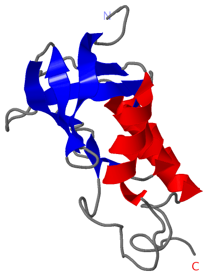
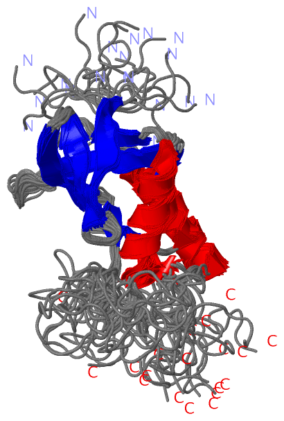
NMR Structure
No.ModelResidues
11, 2, 3, 4, 5, 6, 7, 8, 9, 10, 11, 12, 13, 14, 15, 16, 17, 18, 19, 20Lys A:116 -Pro A:117

 Sequence-Structure Mapping

(-) SAPs(SNPs)/Variants  (0, 0)

(no "SAP(SNP)/Variant" information available for 2M00)

(-) PROSITE Motifs  (3, 3)

NMR Structure (3, 3)
 PROSITEUniProtKBPDB
No.IDACDescriptionIDLocationCountLocation
1TNASE_3PS50830 Thermonuclease domain profile.NUC_STAAW87-221  1A:8-142
2TNASE_1PS01123 Thermonuclease family signature 1.NUC_STAAW98-122  1A:19-42
3TNASE_2PS01284 Thermonuclease family signature 2.NUC_STAAW162-172  1A:83-93
NMR Structure * (3, 3)
 PROSITEUniProtKBPDB
No.IDACDescriptionIDLocationCountLocation
1TNASE_3PS50830 Thermonuclease domain profile.NUC_STAAW87-221  1A:8-142
2TNASE_1PS01123 Thermonuclease family signature 1.NUC_STAAW98-122  1A:19-42
3TNASE_2PS01284 Thermonuclease family signature 2.NUC_STAAW162-172  1A:83-93

(-) Exons   (0, 0)

(no "Exon" information available for 2M00)

(-) Sequences/Alignments

NMR Structure
   Reformat: Number of residues per line =  ('0' or empty: single-line sequence representation)
  Number of residues per labelling interval =   
  UniProt sequence: complete  aligned part    
   Show mapping: SCOP domains CATH domains Pfam domains Secondary structure (by author)
SAPs(SNPs) PROSITE motifs Exons
(details for a mapped element are shown in a popup box when the mouse pointer rests over it)
Chain A from PDB  Type:PROTEIN  Length:149
 aligned with NUC_STAAW | Q8NXI6 from UniProtKB/Swiss-Prot  Length:228

    Alignment length:149
                                    89        99       109       119       129       139       149       159       169       179       189       199       209       219         
            NUC_STAAW    80 ATSTKKLHKEPATLIKAIDGDTVKLMYKGQPMTFRLLLVDTPETKHPKKGVEKYGPEASAFTKKMVENAKKIEVEFDKGQRTDKYGRGLAYIYADGKMVNEALVRQGLAKVAYVYKPNNTHEQLLRKSEAQAKKEKLNIWSEDNADSGQ 228
               SCOP domains d2m00a_ A: Staphylococcal nuclease                                                                                                                    SCOP domains
               CATH domains ----------------------------------------------------------------------------------------------------------------------------------------------------- CATH domains
               Pfam domains ----------------------------------------------------------------------------------------------------------------------------------------------------- Pfam domains
         Sec.struct. author .......eeeeeeeeee....eeeeee..eeeeee...ee...............hhhhhhhhhhhhhh..eeeee...........eeeeeee..eehhhhhhhh...ee.........hhhhhhhhhhhhhhh.............. Sec.struct. author
                 SAPs(SNPs) ----------------------------------------------------------------------------------------------------------------------------------------------------- SAPs(SNPs)
                PROSITE (1) -------TNASE_3  PDB: A:8-142 UniProt: 87-221                                                                                                  ------- PROSITE (1)
                PROSITE (2) ------------------TNASE_1  PDB: A:19-42    ---------------------------------------TNASE_2    -------------------------------------------------------- PROSITE (2)
                 Transcript ----------------------------------------------------------------------------------------------------------------------------------------------------- Transcript
                 2m00 A   1 ATSTKKLHKEPATLIKAIDGDTVKLMYKGQPMTFRLLLVDTPSTKHPKKGVEKYGPEASAFTKKMVENAKKIEVEFDKGQRTDKYGRGLAYIYADGKMVNEALVRQGLAKVAYVYKPNNTHEQLLRKSEAQAKKEKLNIWSEDNADSGQ 149
                                    10        20        30        40        50        60        70        80        90       100       110       120       130       140         

   Legend:   → Mismatch (orange background)
  - → Gap (green background, '-', border residues have a numbering label)
    → Modified Residue (blue background, lower-case, 'x' indicates undefined single-letter code, labelled with number + name)
  x → Chemical Group (purple background, 'x', labelled with number + name, e.g. ACE or NH2)
  extra numbering lines below/above indicate numbering irregularities and modified residue names etc., number ends below/above '|'

 Classification and Annotation

(-) SCOP Domains  (1, 1)

NMR Structure

(-) CATH Domains  (0, 0)

(no "CATH Domain" information available for 2M00)

(-) Pfam Domains  (0, 0)

(no "Pfam Domain" information available for 2M00)

(-) Gene Ontology  (7, 7)

NMR Structure(hide GO term definitions)
Chain A   (NUC_STAAW | Q8NXI6)
molecular function
    GO:0004519    endonuclease activity    Catalysis of the hydrolysis of ester linkages within nucleic acids by creating internal breaks.
    GO:0016787    hydrolase activity    Catalysis of the hydrolysis of various bonds, e.g. C-O, C-N, C-C, phosphoric anhydride bonds, etc. Hydrolase is the systematic name for any enzyme of EC class 3.
    GO:0046872    metal ion binding    Interacting selectively and non-covalently with any metal ion.
    GO:0004518    nuclease activity    Catalysis of the hydrolysis of ester linkages within nucleic acids.
    GO:0003676    nucleic acid binding    Interacting selectively and non-covalently with any nucleic acid.
biological process
    GO:0090305    nucleic acid phosphodiester bond hydrolysis    The nucleic acid metabolic process in which the phosphodiester bonds between nucleotides are cleaved by hydrolysis.
cellular component
    GO:0005576    extracellular region    The space external to the outermost structure of a cell. For cells without external protective or external encapsulating structures this refers to space outside of the plasma membrane. This term covers the host cell environment outside an intracellular parasite.

 Visualization

(-) Interactive Views

NMR Structure
  Complete Structure
    Jena3D(integrated viewing of ligand, site, SAP, PROSITE, SCOP information)
    WebMol | AstexViewer[tm]@PDBe
(Java Applets, require no local installation except for Java; loading may be slow)
    STRAP
(Java WebStart application, automatic local installation, requires Java; full application with system access!)
    RasMol
(require local installation)
    Molscript (VRML)
(requires installation of a VRML viewer; select preferred view via VRML and generate a mono or stereo PDF format file)
 
  Ligands, Modified Residues, Ions
(no "Ligands, Modified Residues, Ions" information available for 2m00)
 
  Sites
(no "Sites" information available for 2m00)
 
  Cis Peptide Bonds
    Lys A:116 - Pro A:117   [ RasMol ]  
 

(-) Still Images

Jmol
  protein: cartoon or spacefill or dots and stick; nucleic acid: cartoon and stick; ligands: spacefill; active site: stick
Molscript
  protein, nucleic acid: cartoon; ligands: spacefill; active site: ball and stick

 Databases and Analysis Tools

(-) Databases

Access by PDB/NDB ID
  2m00
    Family and Domain InformationProDom | SYSTERS
    General Structural InformationGlycoscienceDB | MMDB | NDB | OCA | PDB | PDBe | PDBj | PDBsum | PDBWiki | PQS | PROTEOPEDIA
    Orientation in MembranesOPM
    Protein SurfaceSURFACE
    Secondary StructureDSSP (structure derived) | HSSP (homology derived)
    Structural GenomicsGeneCensus
    Structural NeighboursCE | VAST
    Structure ClassificationCATH | Dali | SCOP
    Validation and Original DataBMRB Data View | BMRB Restraints Grid | EDS | PROCHECK | RECOORD | WHAT_CHECK
 
Access by UniProt ID/Accession number
  NUC_STAAW | Q8NXI6
    Comparative Protein Structure ModelsModBase
    Genomic InformationEnsembl
    Protein-protein InteractionDIP
    Sequence, Family and Domain InformationInterPro | Pfam | SMART | UniProtKB/SwissProt
 
Access by Enzyme Classificator   (EC Number)
  3.1.31.1
    General Enzyme InformationBRENDA | EC-PDB | Enzyme | IntEnz
    PathwayKEGG | MetaCyc
 
Access by Disease Identifier   (MIM ID)
  (no 'MIM ID' available)
    Disease InformationOMIM
 
Access by GenAge ID
  (no 'GenAge ID' available)
    Age Related InformationGenAge

(-) Analysis Tools

Access by PDB/NDB ID
    Domain InformationXDom
    Interatomic Contacts of Structural UnitsCSU
    Ligand-protein ContactsLPC
    Protein CavitiescastP
    Sequence and Secondary StructurePDBCartoon
    Structure AlignmentSTRAP(Java WebStart application, automatic local installation, requires Java; full application with system access!)
    Structure and Sequence BrowserSTING
 
Access by UniProt ID/Accession number
  NUC_STAAW | Q8NXI6
    Protein Disorder PredictionDisEMBL | FoldIndex | GLOBPLOT (for more information see DisProt)

 Related Entries

(-) Entries Sharing at Least One Protein Chain (UniProt ID)

UniProtKB/Swiss-Prot
        NUC_STAAW | Q8NXI62gsi 2lkv 2oxp 2pqe 2pw5 2pw7 2pyk 2pzt 2pzu 2pzw 3d4w 3d8g 3mvv 3qoj 3qol 3qon 3r3o

(-) Related Entries Specified in the PDB File

(no "Related Entries Specified in the PDB File" available for 2M00)