| molecular function |
|---|
| | GO:1990380 | | Lys48-specific deubiquitinase activity | | Hydrolysis of Lys48-linked ubiquitin unit(s) from a ubiquitinated protein. |
| | GO:0008234 | | cysteine-type peptidase activity | | Catalysis of the hydrolysis of peptide bonds in a polypeptide chain by a mechanism in which the sulfhydryl group of a cysteine residue at the active center acts as a nucleophile. |
| | GO:0016787 | | hydrolase activity | | Catalysis of the hydrolysis of various bonds, e.g. C-O, C-N, C-C, phosphoric anhydride bonds, etc. Hydrolase is the systematic name for any enzyme of EC class 3. |
| | GO:0046872 | | metal ion binding | | Interacting selectively and non-covalently with any metal ion. |
| | GO:0008233 | | peptidase activity | | Catalysis of the hydrolysis of a peptide bond. A peptide bond is a covalent bond formed when the carbon atom from the carboxyl group of one amino acid shares electrons with the nitrogen atom from the amino group of a second amino acid. |
| | GO:0004843 | | thiol-dependent ubiquitin-specific protease activity | | Catalysis of the thiol-dependent hydrolysis of a peptide bond formed by the C-terminal glycine of ubiquitin and another protein. |
| | GO:0036459 | | thiol-dependent ubiquitinyl hydrolase activity | | Catalysis of the thiol-dependent hydrolysis of an ester, thioester, amide, peptide or isopeptide bond formed by the C-terminal glycine of ubiquitin. |
| | GO:0031625 | | ubiquitin protein ligase binding | | Interacting selectively and non-covalently with a ubiquitin protein ligase enzyme, any of the E3 proteins. |
| | GO:1904265 | | ubiquitin-specific protease activity involved in negative regulation of retrograde protein transport, ER to cytosol | | Any ubiquitin-specific protease activity (deubiquitinase activity) that is involved in negative regulation of retrograde protein transport, ER to cytosol. |
| biological process |
|---|
| | GO:0030968 | | endoplasmic reticulum unfolded protein response | | The series of molecular signals generated as a consequence of the presence of unfolded proteins in the endoplasmic reticulum (ER) or other ER-related stress; results in changes in the regulation of transcription and translation. |
| | GO:1904153 | | negative regulation of retrograde protein transport, ER to cytosol | | Any process that stops, prevents or reduces the frequency, rate or extent of retrograde protein transport, ER to cytosol. |
| | GO:0035871 | | protein K11-linked deubiquitination | | A protein deubiquitination process in which a K11-linked ubiquitin chain, i.e. a polymer of ubiquitin formed by linkages between lysine residues at position 11 of the ubiquitin monomers, is removed from a protein. |
| | GO:1990167 | | protein K27-linked deubiquitination | | A protein deubiquitination process in which a K27-linked ubiquitin chain, i.e. a polymer of ubiquitin formed by linkages between lysine residues at position 27 of the ubiquitin monomers, is removed from a protein. |
| | GO:0035523 | | protein K29-linked deubiquitination | | A protein deubiquitination process in which a K29-linked ubiquitin chain, i.e. a polymer of ubiquitin formed by linkages between lysine residues at position 29 of the ubiquitin monomers, is removed from a protein. |
| | GO:1990168 | | protein K33-linked deubiquitination | | A protein deubiquitination process in which a K33-linked ubiquitin chain, i.e. a polymer of ubiquitin formed by linkages between lysine residues at position 33 of the ubiquitin monomers, is removed from a protein. |
| | GO:0071108 | | protein K48-linked deubiquitination | | A protein deubiquitination process in which a K48-linked ubiquitin chain, i.e. a polymer of ubiquitin formed by linkages between lysine residues at position 48 of the ubiquitin monomers, is removed from a protein. |
| | GO:0070536 | | protein K63-linked deubiquitination | | A protein deubiquitination process in which a K63-linked ubiquitin chain, i.e. a polymer of ubiquitin formed by linkages between lysine residues at position 63 of the ubiquitin monomers, is removed from a protein. |
| | GO:0006508 | | proteolysis | | The hydrolysis of proteins into smaller polypeptides and/or amino acids by cleavage of their peptide bonds. |
| | GO:0006986 | | response to unfolded protein | | Any process that results in a change in state or activity of a cell or an organism (in terms of movement, secretion, enzyme production, gene expression, etc.) as a result of an unfolded protein stimulus. |
| | GO:0030433 | | ubiquitin-dependent ERAD pathway | | The series of steps necessary to target endoplasmic reticulum (ER)-resident proteins for degradation by the cytoplasmic proteasome. Begins with recognition of the ER-resident protein, includes retrotranslocation (dislocation) of the protein from the ER to the cytosol, protein ubiquitination necessary for correct substrate transfer, transport of the protein to the proteasome, and ends with degradation of the protein by the cytoplasmic proteasome. |
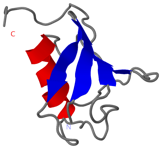
 Description
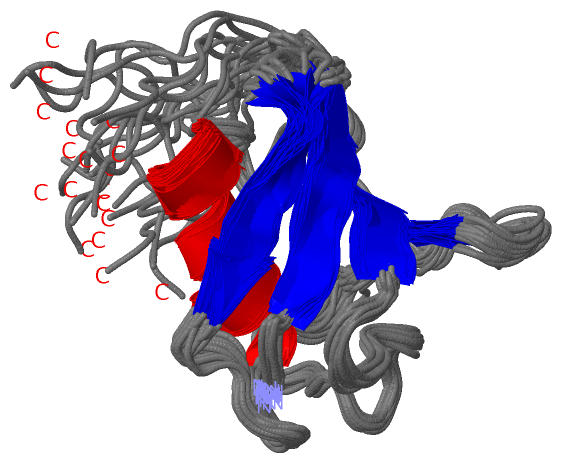