Show PDB file:   
         Plain Text   HTML   (compressed file size)
QuickSearch:   
by PDB,NDB,UniProt,PROSITE Code or Search Term(s)  
(-)NMR Structure - model 1
(-)NMR Structure - all models
collapse expand < >
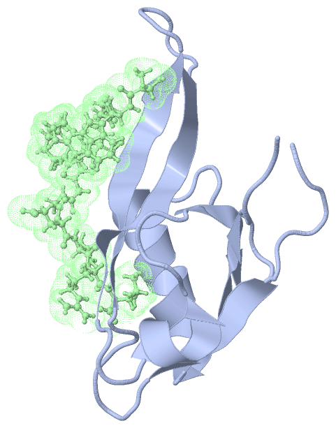
Image NMR Structure - model 1
NMR Structure - model 1  (Jmol Viewer)
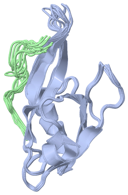
Image NMR Structure - all models
NMR Structure - all models  (Jmol Viewer)

(-) Description

Title :  SOLUTION STRUCTURE OF MAST205-PDZ COMPLEXED WITH THE C-TERMINUS OF A RABIES VIRUS G PROTEIN
 
Authors :  E. Terrien, N. Wolff, F. Cordier, C. Simenel, A. Bernard, M. Lafon, M. De
Date :  04 Nov 09  (Deposition) - 10 Nov 10  (Release) - 10 Nov 10  (Revision)
Method :  SOLUTION NMR
Resolution :  NOT APPLICABLE
Chains :  NMR Structure  :  A,B  (10x)
NMR Structure *:  A,B  (1x)
Keywords :  Pdz Domain, Mast205, Kinase, Pdz Complex And Viral Peptide, Signaling Protein-Peptide Binding Protein Complex (Keyword Search: [Gene Ontology, PubMed, Web (Google))
 
Reference :  E. Terrien, N. Wolff, F. Cordier, C. Simenel, A. Bernard, M. Lafon, M. Delepierre, H. Buc, C. Prehaud, M. Lafage
1H, 13C And 15N Resonance Assignments Of The Pdz Of Microtubule-Associated Serine/Threonine Kinase 205 (Mast205 In Complex With The C-Terminal Motif From The Rabies Virus Glycoprotein
To Be Published
PubMed: search

(-) Compounds

Molecule 1 - MICROTUBULE-ASSOCIATED SERINE/THREONINE-PROTEIN KINASE 2
    ChainsA
    EngineeredYES
    Expression SystemESCHERICHIA COLI BL21(DE3)
    Expression System PlasmidPDEST15
    Expression System StrainBL21(DE3) STAR
    Expression System Taxid469008
    Expression System Vector TypeVECTOR
    FragmentPDZ DOMAIN
    GeneMAST205
    Organism CommonHUMAN
    Organism ScientificHOMO SAPIENS
    Organism Taxid9606
 
Molecule 2 - C-TERMINAL MOTIF FROM GLYCOPROTEIN
    ChainsB
    EngineeredYES
    Other DetailsSYNTHETIC PEPTIDE
    SyntheticYES

 Structural Features

(-) Chains, Units

  12
NMR Structure (10x)AB
NMR Structure * (1x)AB

Summary Information (see also Sequences/Alignments below)

(-) Ligands, Modified Residues, Ions  (0, 0)

(no "Ligand,Modified Residues,Ions" information available for 2KQF)

(-) Sites  (0, 0)

(no "Site" information available for 2KQF)

(-) SS Bonds  (0, 0)

(no "SS Bond" information available for 2KQF)

(-) Cis Peptide Bonds  (0, 0)

(no "Cis Peptide Bond" information available for 2KQF)

 Sequence-Structure Mapping

(-) SAPs(SNPs)/Variants  (0, 0)

(no "SAP(SNP)/Variant" information available for 2KQF)

(-) PROSITE Motifs  (1, 1)

NMR Structure (1, 1)
 PROSITEUniProtKBPDB
No.IDACDescriptionIDLocationCountLocation
1PDZPS50106 PDZ domain profile.MAST2_HUMAN1104-1192  1A:7-95
NMR Structure * (1, 1)
 PROSITEUniProtKBPDB
No.IDACDescriptionIDLocationCountLocation
1PDZPS50106 PDZ domain profile.MAST2_HUMAN1104-1192  1A:7-95

(-) Exons   (0, 0)

(no "Exon" information available for 2KQF)

(-) Sequences/Alignments

NMR Structure
   Reformat: Number of residues per line =  ('0' or empty: single-line sequence representation)
  Number of residues per labelling interval =   
  UniProt sequence: complete  aligned part    
   Show mapping: SCOP domains CATH domains Pfam domains Secondary structure (by author)
SAPs(SNPs) PROSITE motifs Exons
(details for a mapped element are shown in a popup box when the mouse pointer rests over it)
Chain A from PDB  Type:PROTEIN  Length:96
 aligned with MAST2_HUMAN | Q6P0Q8 from UniProtKB/Swiss-Prot  Length:1798

    Alignment length:96
                                  1107      1117      1127      1137      1147      1157      1167      1177      1187      
         MAST2_HUMAN   1098 LGSMRPPIIIHRAGKKYGFTLRAIRVYMGDSDVYTVHHMVWHVEDGGPASEAGLRQGDLITHVNGEPVHGLVHTEVVELILKSGNKVAISTTPLEN 1193
               SCOP domains ------------------------------------------------------------------------------------------------ SCOP domains
               CATH domains ------------------------------------------------------------------------------------------------ CATH domains
               Pfam domains -------PDZ-2kqfA01 A:8-91                                                                  ----- Pfam domains
         Sec.struct. author ......eeeee.......eeeeeeeee......eeeeeeeeee....hhhhhh.....eeeee........hhhhhhhhhhhhh.eeeeee..... Sec.struct. author
                 SAPs(SNPs) ------------------------------------------------------------------------------------------------ SAPs(SNPs)
                    PROSITE ------PDZ  PDB: A:7-95 UniProt: 1104-1192                                                      - PROSITE
                 Transcript ------------------------------------------------------------------------------------------------ Transcript
                2kqf A    1 GGSMRPPIIIHRAGKKYGFTLRAIRVYMGDSDVYTVHHMVWHVEDGGPASEAGLRQGDLITHVNGEPVHGLVHTEVVELILKSGNKVAISTTPLEN   96
                                    10        20        30        40        50        60        70        80        90      

Chain B from PDB  Type:PROTEIN  Length:13
 aligned with Q8JJY9_9RHAB | Q8JJY9 from UniProtKB/TrEMBL  Length:524

    Alignment length:13
                                   521   
        Q8JJY9_9RHAB    512 SWESHKSGGETRL  524
               SCOP domains ------------- SCOP domains
               CATH domains ------------- CATH domains
               Pfam domains ------------- Pfam domains
         Sec.struct. author ............. Sec.struct. author
                 SAPs(SNPs) ------------- SAPs(SNPs)
                    PROSITE ------------- PROSITE
                 Transcript ------------- Transcript
                2kqf B  101 SWESHKSGGETRL  113
                                   110   

   Legend:   → Mismatch (orange background)
  - → Gap (green background, '-', border residues have a numbering label)
    → Modified Residue (blue background, lower-case, 'x' indicates undefined single-letter code, labelled with number + name)
  x → Chemical Group (purple background, 'x', labelled with number + name, e.g. ACE or NH2)
  extra numbering lines below/above indicate numbering irregularities and modified residue names etc., number ends below/above '|'

 Classification and Annotation

(-) SCOP Domains  (0, 0)

(no "SCOP Domain" information available for 2KQF)

(-) CATH Domains  (0, 0)

(no "CATH Domain" information available for 2KQF)

(-) Pfam Domains  (1, 1)

NMR Structure
(-)
Clan: PDZ-like (184)

(-) Gene Ontology  (25, 26)

NMR Structure(hide GO term definitions)
Chain A   (MAST2_HUMAN | Q6P0Q8)
molecular function
    GO:0005524    ATP binding    Interacting selectively and non-covalently with ATP, adenosine 5'-triphosphate, a universally important coenzyme and enzyme regulator.
    GO:0016301    kinase activity    Catalysis of the transfer of a phosphate group, usually from ATP, to a substrate molecule.
    GO:0000287    magnesium ion binding    Interacting selectively and non-covalently with magnesium (Mg) ions.
    GO:0046872    metal ion binding    Interacting selectively and non-covalently with any metal ion.
    GO:0008017    microtubule binding    Interacting selectively and non-covalently with microtubules, filaments composed of tubulin monomers.
    GO:0000166    nucleotide binding    Interacting selectively and non-covalently with a nucleotide, any compound consisting of a nucleoside that is esterified with (ortho)phosphate or an oligophosphate at any hydroxyl group on the ribose or deoxyribose.
    GO:0019902    phosphatase binding    Interacting selectively and non-covalently with any phosphatase.
    GO:0005515    protein binding    Interacting selectively and non-covalently with any protein or protein complex (a complex of two or more proteins that may include other nonprotein molecules).
    GO:0004672    protein kinase activity    Catalysis of the phosphorylation of an amino acid residue in a protein, usually according to the reaction: a protein + ATP = a phosphoprotein + ADP.
    GO:0004674    protein serine/threonine kinase activity    Catalysis of the reactions: ATP + protein serine = ADP + protein serine phosphate, and ATP + protein threonine = ADP + protein threonine phosphate.
    GO:0016740    transferase activity    Catalysis of the transfer of a group, e.g. a methyl group, glycosyl group, acyl group, phosphorus-containing, or other groups, from one compound (generally regarded as the donor) to another compound (generally regarded as the acceptor). Transferase is the systematic name for any enzyme of EC class 2.
biological process
    GO:0007010    cytoskeleton organization    A process that is carried out at the cellular level which results in the assembly, arrangement of constituent parts, or disassembly of cytoskeletal structures.
    GO:0035556    intracellular signal transduction    The process in which a signal is passed on to downstream components within the cell, which become activated themselves to further propagate the signal and finally trigger a change in the function or state of the cell.
    GO:0018105    peptidyl-serine phosphorylation    The phosphorylation of peptidyl-serine to form peptidyl-O-phospho-L-serine.
    GO:0016310    phosphorylation    The process of introducing a phosphate group into a molecule, usually with the formation of a phosphoric ester, a phosphoric anhydride or a phosphoric amide.
    GO:0006468    protein phosphorylation    The process of introducing a phosphate group on to a protein.
    GO:0045075    regulation of interleukin-12 biosynthetic process    Any process that modulates the frequency, rate or extent of the chemical reactions and pathways resulting in the formation of interleukin-12.
    GO:0048515    spermatid differentiation    The process whose specific outcome is the progression of a spermatid over time, from initial commitment of the cell to a specific fate, to the fully functional differentiated cell.
cellular component
    GO:0005737    cytoplasm    All of the contents of a cell excluding the plasma membrane and nucleus, but including other subcellular structures.
    GO:0005856    cytoskeleton    Any of the various filamentous elements that form the internal framework of cells, and typically remain after treatment of the cells with mild detergent to remove membrane constituents and soluble components of the cytoplasm. The term embraces intermediate filaments, microfilaments, microtubules, the microtrabecular lattice, and other structures characterized by a polymeric filamentous nature and long-range order within the cell. The various elements of the cytoskeleton not only serve in the maintenance of cellular shape but also have roles in other cellular functions, including cellular movement, cell division, endocytosis, and movement of organelles.
    GO:0016020    membrane    A lipid bilayer along with all the proteins and protein complexes embedded in it an attached to it.
    GO:0015630    microtubule cytoskeleton    The part of the cytoskeleton (the internal framework of a cell) composed of microtubules and associated proteins.
    GO:0005886    plasma membrane    The membrane surrounding a cell that separates the cell from its external environment. It consists of a phospholipid bilayer and associated proteins.

Chain B   (Q8JJY9_9RHAB | Q8JJY9)
cellular component
    GO:0016021    integral component of membrane    The component of a membrane consisting of the gene products and protein complexes having at least some part of their peptide sequence embedded in the hydrophobic region of the membrane.
    GO:0016020    membrane    A lipid bilayer along with all the proteins and protein complexes embedded in it an attached to it.
    GO:0019031    viral envelope    The lipid bilayer of a virion that surrounds the protein capsid. May also contain glycoproteins.

 Visualization

(-) Interactive Views

NMR Structure
  Complete Structure
    Jena3D(integrated viewing of ligand, site, SAP, PROSITE, SCOP information)
    WebMol | AstexViewer[tm]@PDBe
(Java Applets, require no local installation except for Java; loading may be slow)
    STRAP
(Java WebStart application, automatic local installation, requires Java; full application with system access!)
    RasMol
(require local installation)
    Molscript (VRML)
(requires installation of a VRML viewer; select preferred view via VRML and generate a mono or stereo PDF format file)
 
  Ligands, Modified Residues, Ions
(no "Ligands, Modified Residues, Ions" information available for 2kqf)
 
  Sites
(no "Sites" information available for 2kqf)
 
  Cis Peptide Bonds
(no "Cis Peptide Bonds" information available for 2kqf)
 

(-) Still Images

Jmol
  protein: cartoon or spacefill or dots and stick; nucleic acid: cartoon and stick; ligands: spacefill; active site: stick
Molscript
  protein, nucleic acid: cartoon; ligands: spacefill; active site: ball and stick

 Databases and Analysis Tools

(-) Databases

Access by PDB/NDB ID
  2kqf
    Family and Domain InformationProDom | SYSTERS
    General Structural InformationGlycoscienceDB | MMDB | NDB | OCA | PDB | PDBe | PDBj | PDBsum | PDBWiki | PQS | PROTEOPEDIA
    Orientation in MembranesOPM
    Protein SurfaceSURFACE
    Secondary StructureDSSP (structure derived) | HSSP (homology derived)
    Structural GenomicsGeneCensus
    Structural NeighboursCE | VAST
    Structure ClassificationCATH | Dali | SCOP
    Validation and Original DataBMRB Data View | BMRB Restraints Grid | EDS | PROCHECK | RECOORD | WHAT_CHECK
 
Access by UniProt ID/Accession number
  MAST2_HUMAN | Q6P0Q8
    Comparative Protein Structure ModelsModBase
    Genomic InformationEnsembl
    Protein-protein InteractionDIP
    Sequence, Family and Domain InformationInterPro | Pfam | SMART | UniProtKB/SwissProt
  Q8JJY9_9RHAB | Q8JJY9
    Comparative Protein Structure ModelsModBase
    Genomic InformationEnsembl
    Protein-protein InteractionDIP
    Sequence, Family and Domain InformationInterPro | Pfam | SMART | UniProtKB/TrEMBL
 
Access by Enzyme Classificator   (EC Number)
  (no 'Enzyme Classificator' available)
    General Enzyme InformationBRENDA | EC-PDB | Enzyme | IntEnz
    PathwayKEGG | MetaCyc
 
Access by Disease Identifier   (MIM ID)
  (no 'MIM ID' available)
    Disease InformationOMIM
 
Access by GenAge ID
  (no 'GenAge ID' available)
    Age Related InformationGenAge

(-) Analysis Tools

Access by PDB/NDB ID
    Domain InformationXDom
    Interatomic Contacts of Structural UnitsCSU
    Ligand-protein ContactsLPC
    Protein CavitiescastP
    Sequence and Secondary StructurePDBCartoon
    Structure AlignmentSTRAP(Java WebStart application, automatic local installation, requires Java; full application with system access!)
    Structure and Sequence BrowserSTING
 
Access by UniProt ID/Accession number
  MAST2_HUMAN | Q6P0Q8
    Protein Disorder PredictionDisEMBL | FoldIndex | GLOBPLOT (for more information see DisProt)
  Q8JJY9_9RHAB | Q8JJY9
    Protein Disorder PredictionDisEMBL | FoldIndex | GLOBPLOT (for more information see DisProt)

 Related Entries

(-) Entries Sharing at Least One Protein Chain (UniProt ID)

UniProtKB/Swiss-Prot
        MAST2_HUMAN | Q6P0Q82kyl

(-) Related Entries Specified in the PDB File

(no "Related Entries Specified in the PDB File" available for 2KQF)