|
|
|
|
 Description
|
|
Compounds
|
||||||||||||||||||||||||||||||||||||||||||||
Chains, Units
Summary Information (see also Sequences/Alignments below) |
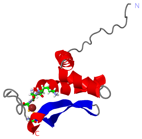
Ligands, Modified Residues, Ions (1, 1)
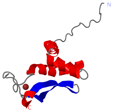
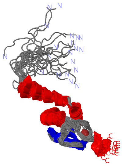
NMR Structure (1, 1)
|
Sites (1, 1)
NMR Structure (1, 1)
|
SS Bonds (0, 0)| (no "SS Bond" information available for 2K7R) |
Cis Peptide Bonds (0, 0)| (no "Cis Peptide Bond" information available for 2K7R) |
SAPs(SNPs)/Variants (0, 0)| (no "SAP(SNP)/Variant" information available for 2K7R) |
PROSITE Motifs (0, 0)| (no "PROSITE Motif" information available for 2K7R) |
Exons (0, 0)| (no "Exon" information available for 2K7R) |
Sequences/Alignments
NMR StructureChain A from PDB Type:PROTEIN Length:106 aligned with DNAI_BACSU | P06567 from UniProtKB/Swiss-Prot Length:311 Alignment length:106 10 20 30 40 50 60 70 80 90 100 DNAI_BACSU 1 MEPIGRSLQGVTGRPDFQKRLEQMKEKVMKDQDVQAFLKENEEVIDQKMIEKSLNKLYEYIEQSKNCSYCSEDENCNNLLEGYHPKLVVNGRSIDIEYYECPVKRK 106 SCOP domains ---------------------------------------------------------------------------------------------------------- SCOP domains CATH domains ---------------------------------------------------------------------------------------------------------- CATH domains Pfam domains DnaI_N-2k7rA01 A:1-98 -------- Pfam domains SAPs(SNPs) ---------------------------------------------------------------------------------------------------------- SAPs(SNPs) PROSITE ---------------------------------------------------------------------------------------------------------- PROSITE Transcript ---------------------------------------------------------------------------------------------------------- Transcript 2k7r A 1 MEPIGRSLQGVTGRPDFQKRLEQMKEKVMKDQDVQAFLKENEEVIDQKMIEKSLNKLYEYIEQSKNCSYCSEDENCNNLLEGYHPKLVVNGRSIDIEYYECPVKRK 106 10 20 30 40 50 60 70 80 90 100
|
||||||||||||||||||||
SCOP Domains (0, 0)| (no "SCOP Domain" information available for 2K7R) |
CATH Domains (0, 0)| (no "CATH Domain" information available for 2K7R) |
Pfam Domains (1, 1)
NMR Structure
|
Gene Ontology (7, 7)|
NMR Structure(hide GO term definitions) Chain A (DNAI_BACSU | P06567)
|
||||||||||||||||||||||||||||||||||||||||||||||||||||||||||||
Interactive Views
|
||||||||||||||||||||||||||||||||||||||||||||||||||||||||||||||||||||||||||||||||||||||||||||||||||||||||||||||||||||||
Still Images
|
||||||||||||||||
Databases
|
||||||||||||||||||||||||||||||||||||||||||||||||||||||||||||||||||||||||||||||||||||||||||||||||||||||||||||||||||||||||||||||||||||||||||||||||||||||||||||||||
Analysis Tools
|
|||||||||||||||||||||||||||||||||||||||||||||||||||||||||||||
Entries Sharing at Least One Protein Chain (UniProt ID)
Related Entries Specified in the PDB File
|
|