Show PDB file:   
         Plain Text   HTML   (compressed file size)
QuickSearch:   
by PDB,NDB,UniProt,PROSITE Code or Search Term(s)  
(-)NMR Structure - manually
(-)NMR Structure - model 1
(-)NMR Structure - all models
collapse expand < >
Image NMR Structure - manually
NMR Structure - manually  (Jmol Viewer)
Image NMR Structure - model 1
NMR Structure - model 1  (Jmol Viewer)
Image NMR Structure - all models
NMR Structure - all models  (Jmol Viewer)

(-) Description

Title :  SOLUTION STRUCTURE OF THE SECOND CLIP DOMAIN IN PAP2
 
Authors :  R. D. Huang, Z. Q. Lv, H. E. Dai, D. V. Velde, O. Prakash, H. B. Jiang
Date :  02 Oct 06  (Deposition) - 16 Oct 07  (Release) - 24 Feb 09  (Revision)
Method :  SOLUTION NMR
Resolution :  NOT APPLICABLE
Chains :  NMR Structure  :  A  (20x)
Keywords :  Beta-Sheet, Double Helix, Hydrolase (Keyword Search: [Gene Ontology, PubMed, Web (Google))
 
Reference :  R. D. Huang, Z. Q. Lv, H. E. Dai, D. V. Velde, O. Prakash, H. B. Jiang
Solution Structure Of Clip Domain In Pap2
To Be Published
PubMed: search
(for further references see the PDB file header)

(-) Compounds

Molecule 1 - PROPHENOLOXIDASE ACTIVATING PROTEINASE-2
    ChainsA
    EngineeredYES
    Expression SystemESCHERICHIA COLI
    Expression System PlasmidPQE60
    Expression System StrainM15
    Expression System Taxid562
    Expression System Vector TypePLASMID
    FragmentSECOND CLIP DOMAIN
    GenePAP-2
    Organism CommonTOBACCO HORNWORM
    Organism ScientificMANDUCA SEXTA
    Organism Taxid7130

 Structural Features

(-) Chains, Units

  
NMR Structure (20x)

Summary Information (see also Sequences/Alignments below)

(-) Ligands, Modified Residues, Ions  (0, 0)

(no "Ligand,Modified Residues,Ions" information available for 2IKE)

(-) Sites  (0, 0)

(no "Site" information available for 2IKE)
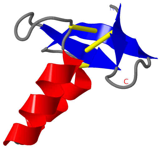
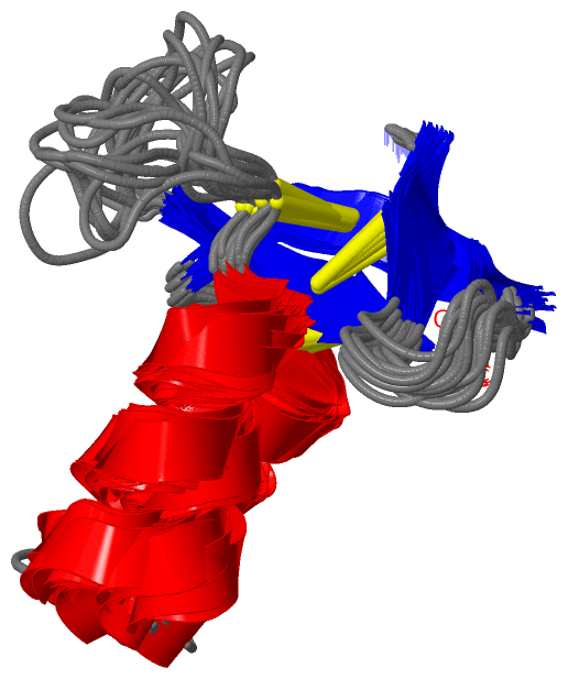
(-) SS Bonds  (3, 3)

NMR Structure
No.Residues
1A:73 -A:122
2A:83 -A:113
3A:89 -A:123

(-) Cis Peptide Bonds  (0, 0)

(no "Cis Peptide Bond" information available for 2IKE)

 Sequence-Structure Mapping

(-) SAPs(SNPs)/Variants  (0, 0)

(no "SAP(SNP)/Variant" information available for 2IKE)

(-) PROSITE Motifs  (0, 0)

(no "PROSITE Motif" information available for 2IKE)

(-) Exons   (0, 0)

(no "Exon" information available for 2IKE)

(-) Sequences/Alignments

NMR Structure
   Reformat: Number of residues per line =  ('0' or empty: single-line sequence representation)
  Number of residues per labelling interval =   
  UniProt sequence: complete  aligned part    
   Show mapping: SCOP domains CATH domains Pfam domains Secondary structure (by author)
SAPs(SNPs) PROSITE motifs Exons
(details for a mapped element are shown in a popup box when the mouse pointer rests over it)
Chain A from PDB  Type:PROTEIN  Length:54
 aligned with Q2FAY5_MANSE | Q2FAY5 from UniProtKB/TrEMBL  Length:441

    Alignment length:54
                                    89        99       109       119       129    
         Q2FAY5_MANSE    80 LSCLTPDNKPGKCVNIKKCTHLAEIEEDPIGEDETTYLKNSVCAGPEDNSVCCG 133
               SCOP domains ------------------------------------------------------ SCOP domains
               CATH domains ------------------------------------------------------ CATH domains
               Pfam domains ------------------------------------------------------ Pfam domains
         Sec.struct. author .eee.....eeeeee...hhhhhhhhhh..hhhhhhhhhhh........eeee. Sec.struct. author
                 SAPs(SNPs) ------------------------------------------------------ SAPs(SNPs)
                    PROSITE ------------------------------------------------------ PROSITE
                 Transcript ------------------------------------------------------ Transcript
                 2ike A  71 LSCLTPDNKPGKCVNIKKCTHLAEIEEDPIGEDETTYLKNSVCAGPEDNSVCCG 124
                                    80        90       100       110       120    

Chain A from PDB  Type:PROTEIN  Length:54
 aligned with Q8I917_MANSE | Q8I917 from UniProtKB/TrEMBL  Length:441

    Alignment length:54
                                    89        99       109       119       129    
         Q8I917_MANSE    80 LSCLTPDNKPGKCVNIKKCTHLAEIEEDPIGEDETTYLKNSVCAGPEDNSVCCG 133
               SCOP domains ------------------------------------------------------ SCOP domains
               CATH domains ------------------------------------------------------ CATH domains
               Pfam domains ------------------------------------------------------ Pfam domains
         Sec.struct. author .eee.....eeeeee...hhhhhhhhhh..hhhhhhhhhhh........eeee. Sec.struct. author
                 SAPs(SNPs) ------------------------------------------------------ SAPs(SNPs)
                    PROSITE ------------------------------------------------------ PROSITE
                 Transcript ------------------------------------------------------ Transcript
                 2ike A  71 LSCLTPDNKPGKCVNIKKCTHLAEIEEDPIGEDETTYLKNSVCAGPEDNSVCCG 124
                                    80        90       100       110       120    

   Legend:   → Mismatch (orange background)
  - → Gap (green background, '-', border residues have a numbering label)
    → Modified Residue (blue background, lower-case, 'x' indicates undefined single-letter code, labelled with number + name)
  x → Chemical Group (purple background, 'x', labelled with number + name, e.g. ACE or NH2)
  extra numbering lines below/above indicate numbering irregularities and modified residue names etc., number ends below/above '|'

 Classification and Annotation

(-) SCOP Domains  (0, 0)

(no "SCOP Domain" information available for 2IKE)

(-) CATH Domains  (0, 0)

(no "CATH Domain" information available for 2IKE)

(-) Pfam Domains  (0, 0)

(no "Pfam Domain" information available for 2IKE)

(-) Gene Ontology  (5, 10)

NMR Structure(hide GO term definitions)
Chain A   (Q2FAY5_MANSE | Q2FAY5)
molecular function
    GO:0016787    hydrolase activity    Catalysis of the hydrolysis of various bonds, e.g. C-O, C-N, C-C, phosphoric anhydride bonds, etc. Hydrolase is the systematic name for any enzyme of EC class 3.
    GO:0008233    peptidase activity    Catalysis of the hydrolysis of a peptide bond. A peptide bond is a covalent bond formed when the carbon atom from the carboxyl group of one amino acid shares electrons with the nitrogen atom from the amino group of a second amino acid.
    GO:0004252    serine-type endopeptidase activity    Catalysis of the hydrolysis of internal, alpha-peptide bonds in a polypeptide chain by a catalytic mechanism that involves a catalytic triad consisting of a serine nucleophile that is activated by a proton relay involving an acidic residue (e.g. aspartate or glutamate) and a basic residue (usually histidine).
    GO:0008236    serine-type peptidase activity    Catalysis of the hydrolysis of peptide bonds in a polypeptide chain by a catalytic mechanism that involves a catalytic triad consisting of a serine nucleophile that is activated by a proton relay involving an acidic residue (e.g. aspartate or glutamate) and a basic residue (usually histidine).
biological process
    GO:0006508    proteolysis    The hydrolysis of proteins into smaller polypeptides and/or amino acids by cleavage of their peptide bonds.

Chain A   (Q8I917_MANSE | Q8I917)
molecular function
    GO:0016787    hydrolase activity    Catalysis of the hydrolysis of various bonds, e.g. C-O, C-N, C-C, phosphoric anhydride bonds, etc. Hydrolase is the systematic name for any enzyme of EC class 3.
    GO:0008233    peptidase activity    Catalysis of the hydrolysis of a peptide bond. A peptide bond is a covalent bond formed when the carbon atom from the carboxyl group of one amino acid shares electrons with the nitrogen atom from the amino group of a second amino acid.
    GO:0004252    serine-type endopeptidase activity    Catalysis of the hydrolysis of internal, alpha-peptide bonds in a polypeptide chain by a catalytic mechanism that involves a catalytic triad consisting of a serine nucleophile that is activated by a proton relay involving an acidic residue (e.g. aspartate or glutamate) and a basic residue (usually histidine).
    GO:0008236    serine-type peptidase activity    Catalysis of the hydrolysis of peptide bonds in a polypeptide chain by a catalytic mechanism that involves a catalytic triad consisting of a serine nucleophile that is activated by a proton relay involving an acidic residue (e.g. aspartate or glutamate) and a basic residue (usually histidine).
biological process
    GO:0006508    proteolysis    The hydrolysis of proteins into smaller polypeptides and/or amino acids by cleavage of their peptide bonds.

 Visualization

(-) Interactive Views

NMR Structure
  Complete Structure
    Jena3D(integrated viewing of ligand, site, SAP, PROSITE, SCOP information)
    WebMol | AstexViewer[tm]@PDBe
(Java Applets, require no local installation except for Java; loading may be slow)
    STRAP
(Java WebStart application, automatic local installation, requires Java; full application with system access!)
    RasMol
(require local installation)
    Molscript (VRML)
(requires installation of a VRML viewer; select preferred view via VRML and generate a mono or stereo PDF format file)
 
  Ligands, Modified Residues, Ions
(no "Ligands, Modified Residues, Ions" information available for 2ike)
 
  Sites
(no "Sites" information available for 2ike)
 
  Cis Peptide Bonds
(no "Cis Peptide Bonds" information available for 2ike)
 

(-) Still Images

Jmol
  protein: cartoon or spacefill or dots and stick; nucleic acid: cartoon and stick; ligands: spacefill; active site: stick
Molscript
  protein, nucleic acid: cartoon; ligands: spacefill; active site: ball and stick

 Databases and Analysis Tools

(-) Databases

Access by PDB/NDB ID
  2ike
    Family and Domain InformationProDom | SYSTERS
    General Structural InformationGlycoscienceDB | MMDB | NDB | OCA | PDB | PDBe | PDBj | PDBsum | PDBWiki | PQS | PROTEOPEDIA
    Orientation in MembranesOPM
    Protein SurfaceSURFACE
    Secondary StructureDSSP (structure derived) | HSSP (homology derived)
    Structural GenomicsGeneCensus
    Structural NeighboursCE | VAST
    Structure ClassificationCATH | Dali | SCOP
    Validation and Original DataBMRB Data View | BMRB Restraints Grid | EDS | PROCHECK | RECOORD | WHAT_CHECK
 
Access by UniProt ID/Accession number
  Q2FAY5_MANSE | Q2FAY5
    Comparative Protein Structure ModelsModBase
    Genomic InformationEnsembl
    Protein-protein InteractionDIP
    Sequence, Family and Domain InformationInterPro | Pfam | SMART | UniProtKB/SwissProt
  Q8I917_MANSE | Q8I917
    Comparative Protein Structure ModelsModBase
    Genomic InformationEnsembl
    Protein-protein InteractionDIP
    Sequence, Family and Domain InformationInterPro | Pfam | SMART | UniProtKB/TrEMBL
 
Access by Enzyme Classificator   (EC Number)
  (no 'Enzyme Classificator' available)
    General Enzyme InformationBRENDA | EC-PDB | Enzyme | IntEnz
    PathwayKEGG | MetaCyc
 
Access by Disease Identifier   (MIM ID)
  (no 'MIM ID' available)
    Disease InformationOMIM
 
Access by GenAge ID
  (no 'GenAge ID' available)
    Age Related InformationGenAge

(-) Analysis Tools

Access by PDB/NDB ID
    Domain InformationXDom
    Interatomic Contacts of Structural UnitsCSU
    Ligand-protein ContactsLPC
    Protein CavitiescastP
    Sequence and Secondary StructurePDBCartoon
    Structure AlignmentSTRAP(Java WebStart application, automatic local installation, requires Java; full application with system access!)
    Structure and Sequence BrowserSTING
 
Access by UniProt ID/Accession number
  Q2FAY5_MANSE | Q2FAY5
    Protein Disorder PredictionDisEMBL | FoldIndex | GLOBPLOT (for more information see DisProt)
  Q8I917_MANSE | Q8I917
    Protein Disorder PredictionDisEMBL | FoldIndex | GLOBPLOT (for more information see DisProt)

 Related Entries

(-) Entries Sharing at Least One Protein Chain (UniProt ID)

UniProtKB/Swiss-Prot
        Q2FAY5_MANSE | Q2FAY52ikd
UniProtKB/TrEMBL
        Q8I917_MANSE | Q8I9172ikd

(-) Related Entries Specified in the PDB File

2ikd