| molecular function |
|---|
| | GO:0016787 | | hydrolase activity | | Catalysis of the hydrolysis of various bonds, e.g. C-O, C-N, C-C, phosphoric anhydride bonds, etc. Hydrolase is the systematic name for any enzyme of EC class 3. |
| | GO:0046872 | | metal ion binding | | Interacting selectively and non-covalently with any metal ion. |
| | GO:0000224 | | peptide-N4-(N-acetyl-beta-glucosaminyl)asparagine amidase activity | | Catalysis of the reaction: 4-N-(N-acetyl-D-glucosaminyl)-protein + H2O = N-acetyl-beta-D-glucosaminylamine + peptide L-aspartate. This reaction is the hydrolysis of an N4-(acetyl-beta-D-glucosaminyl)asparagine residue in which the N-acetyl-D-glucosamine residue may be further glycosylated, to yield a (substituted) N-acetyl-beta-D-glucosaminylamine and the peptide containing an aspartic residue. |
| | GO:0005515 | | protein binding | | Interacting selectively and non-covalently with any protein or protein complex (a complex of two or more proteins that may include other nonprotein molecules). |
| biological process |
|---|
| | GO:0006516 | | glycoprotein catabolic process | | The chemical reactions and pathways resulting in the breakdown of glycoproteins, any protein that contains covalently bound glycose (i.e. monosaccharide) residues; the glycose occurs most commonly as oligosaccharide or fairly small polysaccharide but occasionally as monosaccharide. |
| cellular component |
|---|
| | GO:0005737 | | cytoplasm | | All of the contents of a cell excluding the plasma membrane and nucleus, but including other subcellular structures. |
| | GO:0005634 | | nucleus | | A membrane-bounded organelle of eukaryotic cells in which chromosomes are housed and replicated. In most cells, the nucleus contains all of the cell's chromosomes except the organellar chromosomes, and is the site of RNA synthesis and processing. In some species, or in specialized cell types, RNA metabolism or DNA replication may be absent. |
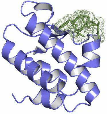
 Description
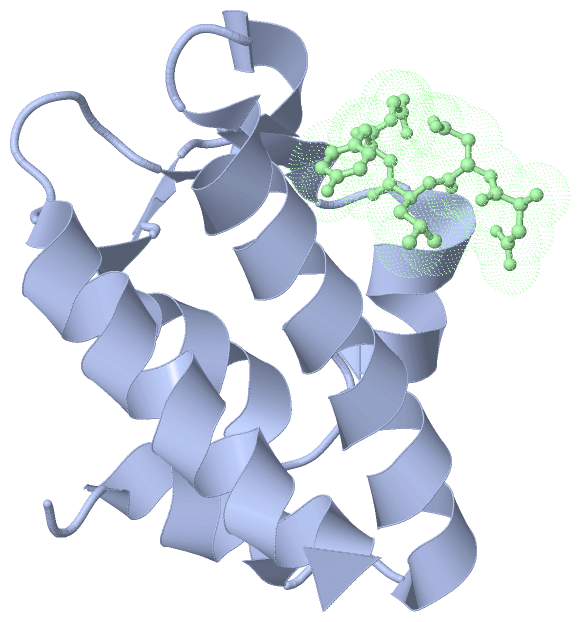