|
|
|
|
 Description
|
|
Compounds
|
||||||||||||||||||||||||||||||||||||||||||||||||||||
Chains, Units
Summary Information (see also Sequences/Alignments below) |
Ligands, Modified Residues, Ions (1, 1)
Asymmetric Unit (1, 1)
|
Sites (1, 1)
Asymmetric Unit (1, 1)
|
SS Bonds (8, 8)
Asymmetric Unit
|
||||||||||||||||||||||||||||||||||||
Cis Peptide Bonds (0, 0)| (no "Cis Peptide Bond" information available for 2H5Z) |
SAPs(SNPs)/Variants (0, 0)| (no "SAP(SNP)/Variant" information available for 2H5Z) |
PROSITE Motifs (2, 4)
Asymmetric Unit (2, 4)
|
||||||||||||||||||||||||||||||||||||||||||||||||||||||||||||||||||||||||||||||||||||||||||||||||
Exons (0, 0)| (no "Exon" information available for 2H5Z) |
Sequences/Alignments
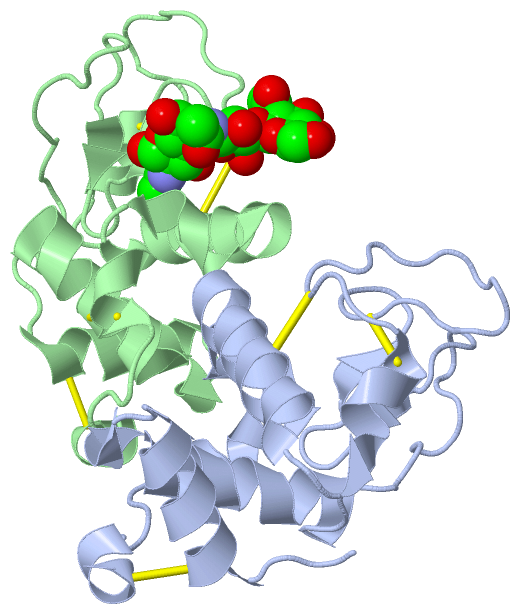
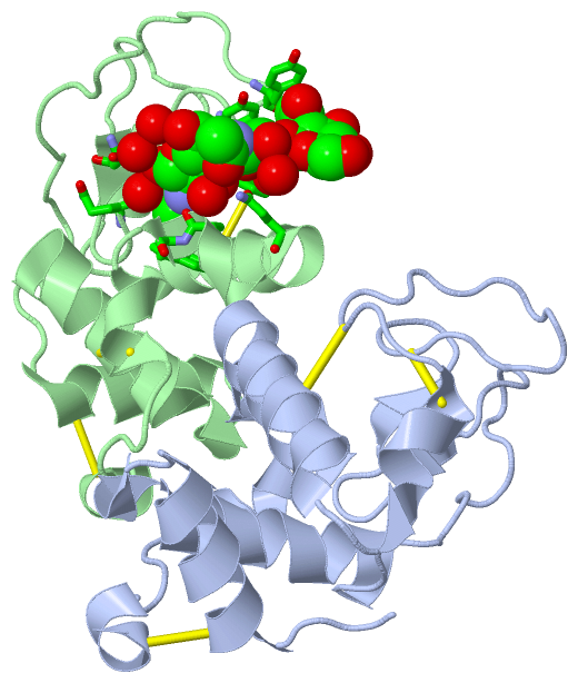
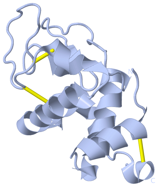
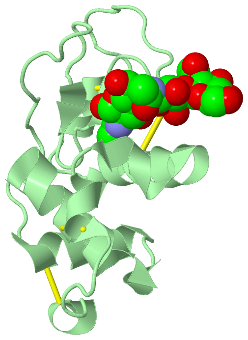
Asymmetric UnitChain A from PDB Type:PROTEIN Length:122 aligned with LYS1_MUSDO | Q7YT16 from UniProtKB/Swiss-Prot Length:141 Alignment length:122 29 39 49 59 69 79 89 99 109 119 129 139 LYS1_MUSDO 20 KTFTRCSLAREMYALGVPKSELPQWTCIAEHESSYRTNVVGPTNSNGSNDYGIFQINNYYWCQPSNGRFSYNECHLSCDALLTDNISNSVTCARKIKSQQGWTAWSTWKYCSGSLPSINDCF 141 SCOP domains d2h5za_ A: automated matches SCOP domains CATH domains 2h5zA00 A:1-122 [code=1.10.530.10, no name defined] CATH domains Pfam domains -------------------------------------------------------------------------------------------------------------------------- Pfam domains SAPs(SNPs) -------------------------------------------------------------------------------------------------------------------------- SAPs(SNPs) PROSITE (1) LACTALBUMIN_LYSOZYME_2 PDB: A:1-122 UniProt: 20-141 PROSITE (1) PROSITE (2) -------------------------------------------------------------------------LACTALBUMIN_LYSOZYM------------------------------ PROSITE (2) Transcript -------------------------------------------------------------------------------------------------------------------------- Transcript 2h5z A 1 KTFTRCSLAREMYALGVPKSELPQWTCIAEHESSYRTNVVGPTNSNGSNDYGIFQINNYYWCQPSNGRFSYNECHLSCDALLTDNISNSVTCARKIKSQQGWTAWSTWKYCSGSLPSINDCF 122 10 20 30 40 50 60 70 80 90 100 110 120 Chain B from PDB Type:PROTEIN Length:122 aligned with LYS1_MUSDO | Q7YT16 from UniProtKB/Swiss-Prot Length:141 Alignment length:122 29 39 49 59 69 79 89 99 109 119 129 139 LYS1_MUSDO 20 KTFTRCSLAREMYALGVPKSELPQWTCIAEHESSYRTNVVGPTNSNGSNDYGIFQINNYYWCQPSNGRFSYNECHLSCDALLTDNISNSVTCARKIKSQQGWTAWSTWKYCSGSLPSINDCF 141 SCOP domains d2h5zb_ B: automated matches SCOP domains CATH domains 2h5zB00 B:1-122 [code=1.10.530.10, no name defined] CATH domains Pfam domains -------------------------------------------------------------------------------------------------------------------------- Pfam domains SAPs(SNPs) -------------------------------------------------------------------------------------------------------------------------- SAPs(SNPs) PROSITE (1) LACTALBUMIN_LYSOZYME_2 PDB: B:1-122 UniProt: 20-141 PROSITE (1) PROSITE (2) -------------------------------------------------------------------------LACTALBUMIN_LYSOZYM------------------------------ PROSITE (2) Transcript -------------------------------------------------------------------------------------------------------------------------- Transcript 2h5z B 1 KTFTRCSLAREMYALGVPKSELPQWTCIAEHESSYRTNVVGPTNSNGSNDYGIFQINNYYWCQPSNGRFSYNECHLSCDALLTDNISNSVTCARKIKSQQGWTAWSTWKYCSGSLPSINDCF 122 10 20 30 40 50 60 70 80 90 100 110 120
|
||||||||||||||||||||
SCOP Domains (1, 2)
Asymmetric Unit
 |
CATH Domains (1, 2)
Asymmetric Unit
|
Pfam Domains (0, 0)| (no "Pfam Domain" information available for 2H5Z) |
Gene Ontology (4, 4)|
Asymmetric Unit(hide GO term definitions) Chain A,B (LYS1_MUSDO | Q7YT16)
|
||||||||||||||||||||||||||||||||||||
Interactive Views
|
|||||||||||||||||||||||||||||||||||||||||||||||||||||||||||||||||||||||||||||||||||||||||||||||||||||||||||||||||||||||||||||||||||||||||||||
Still Images
|
||||||||||||||||
Databases
|
||||||||||||||||||||||||||||||||||||||||||||||||||||||||||||||||||||||||||||||||||||||||||||||||||||||||||||||||||||||||||||||||||||||||||||||||||||||||||||||||
Analysis Tools
|
|||||||||||||||||||||||||||||||||||||||||||||||||||||||||||||
Entries Sharing at Least One Protein Chain (UniProt ID)
Related Entries Specified in the PDB File
|
|