Show PDB file:   
         Plain Text   HTML   (compressed file size)
QuickSearch:   
by PDB,NDB,UniProt,PROSITE Code or Search Term(s)  
(-)Asym./Biol. Unit
(-)Asym./Biol. Unit - sites
collapse expand < >
Image Asym./Biol. Unit
Asym./Biol. Unit  (Jmol Viewer)
Image Asym./Biol. Unit - sites
Asym./Biol. Unit - sites  (Jmol Viewer)

(-) Description

Title :  S.PNEUMONIAE PDF COMPLEXED WITH SB-543668
 
Authors :  K. J. Smith, C. M. Petit, K. Aubart, M. Smyth, E. Mcmanus, J. Jones, A. Fosberry, C. Lewis, M. Lonetto, S. B. Christensen
Date :  29 Jul 05  (Deposition) - 06 Sep 05  (Release) - 24 Feb 09  (Revision)
Method :  X-RAY DIFFRACTION
Resolution :  1.70
Chains :  Asym./Biol. Unit :  A
Keywords :  Hydrolase (Keyword Search: [Gene Ontology, PubMed, Web (Google))
 
Reference :  K. J. Smith, C. M. Petit, K. Aubart, M. Smyth, E. Mcmanus, J. Jones, A. Fosberry, C. Lewis, M. Lonetto, S. B. Christensen
Structural Variation And Inhibitor Binding In Polypeptide Deformylase From Four Different Bacterial Species.
Protein Sci. V. 12 349 2003
PubMed-ID: 12538898  |  Reference-DOI: 10.1110/PS.0229303
(for further references see the PDB file header)

(-) Compounds

Molecule 1 - PEPTIDE DEFORMYLASE
    ChainsA
    EC Number3.5.1.88
    EngineeredYES
    Expression SystemESCHERICHIA COLI
    Expression System Taxid562
    GeneDEF
    Organism ScientificSTREPTOCOCCUS PNEUMONIAE
    Organism Taxid1313
    StrainATCC BAA-255-R6
    SynonymPDF, POLYPEPTIDE DEFORMYLASE

 Structural Features

(-) Chains, Units

  1
Asymmetric/Biological Unit A

Summary Information (see also Sequences/Alignments below)
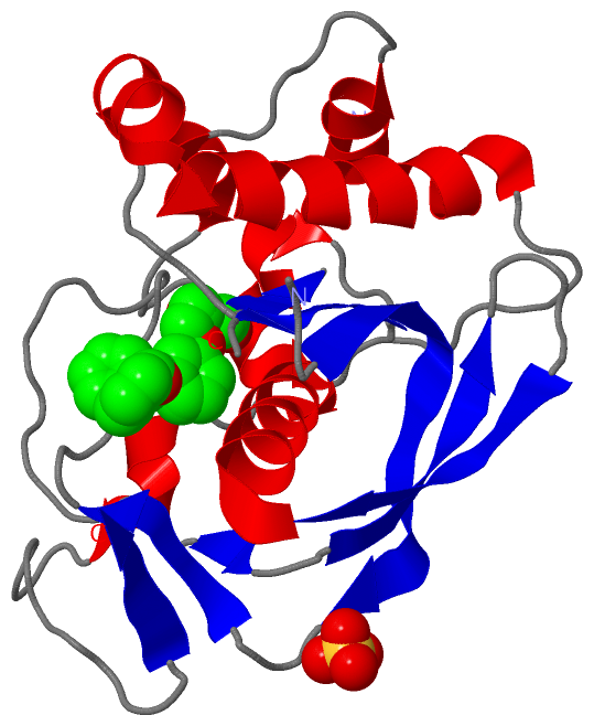
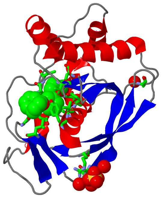
(-) Ligands, Modified Residues, Ions  (3, 3)

Asymmetric/Biological Unit (3, 3)
No.NameCountTypeFull Name
1NI1Ligand/IonNICKEL (II) ION
2SB81Ligand/Ion2-(3-BENZOYLPHENOXY)ETHYL(HYDROXY)FORMAMIDE
3SO41Ligand/IonSULFATE ION

(-) Sites  (3, 3)

Asymmetric Unit (3, 3)
No.NameEvidenceResiduesDescription
1AC1SOFTWAREGLN A:76 , CYS A:130 , HIS A:173 , HIS A:177 , SB8 A:501BINDING SITE FOR RESIDUE NI A 601
2AC2SOFTWARETHR A:34 , SER A:117 , ARG A:146 , HOH A:3031 , HOH A:3088 , HOH A:3141 , HOH A:3176BINDING SITE FOR RESIDUE SO4 A 3001
3AC3SOFTWAREVAL A:70 , GLY A:71 , GLN A:76 , LEU A:123 , GLY A:126 , GLU A:128 , GLY A:129 , CYS A:130 , LEU A:131 , TYR A:166 , VAL A:170 , HIS A:173 , GLU A:174 , HIS A:177 , NI A:601BINDING SITE FOR RESIDUE SB8 A 501

(-) SS Bonds  (0, 0)

(no "SS Bond" information available for 2AIA)

(-) Cis Peptide Bonds  (1, 1)

Asymmetric/Biological Unit
No.Residues
1Phe A:35 -Pro A:36

 Sequence-Structure Mapping

(-) SAPs(SNPs)/Variants  (0, 0)

(no "SAP(SNP)/Variant" information available for 2AIA)

(-) PROSITE Motifs  (0, 0)

(no "PROSITE Motif" information available for 2AIA)

(-) Exons   (0, 0)

(no "Exon" information available for 2AIA)

(-) Sequences/Alignments

Asymmetric/Biological Unit
   Reformat: Number of residues per line =  ('0' or empty: single-line sequence representation)
  Number of residues per labelling interval =   
  UniProt sequence: complete  aligned part    
   Show mapping: SCOP domains CATH domains Pfam domains Secondary structure (by author)
SAPs(SNPs) PROSITE motifs Exons
(details for a mapped element are shown in a popup box when the mouse pointer rests over it)
Chain A from PDB  Type:PROTEIN  Length:192
 aligned with DEF_STRR6 | Q8DP79 from UniProtKB/Swiss-Prot  Length:203

    Alignment length:202
                                    11        21        31        41        51        61        71        81        91       101       111       121       131       141       151       161       171       181       191       201  
            DEF_STRR6     2 SAIERITKAAHLIDMNDIIREGNPTLRTVAEEVTFPLSDQEIILGEKMMQFLKHSQDPVMAEKMGLRGGVGLAAPQLDISKRIIAVLVPNIVEEGETPQEAYDLEAIMYNPKIVSHSVQDAALGEGEGCLSVDRNVPGYVVRHARVTVDYFDKDGEKHRIKLKGYNSIVVQHEIDHINGIMFYDRINEKDPFAVKDGLLILE 203
               SCOP domains d2aiaa_ A: Peptide deformylase                                                                                                                                                                             SCOP domains
               CATH domains 2aiaA00 A:1-203 Peptide Deformylase                                                                                                                                                                        CATH domains
               Pfam domains ---------------------------------------------------------------------------------------------------------------------------------------------------------------------------------------------------------- Pfam domains
         Sec.struct. author hhhhhhh......hhhhh....hhhhhh.........hhhhhhhhhhhhhhhhhhhhhhhhhhhh.....eee.hhhh....eeeeee..----------...eeeeeeeeeeeee...eeee..................ee..eeeeee.....eeeeeehhhhhhhhhhhhhhhh..hhhhhh...........eeee. Sec.struct. author
                 SAPs(SNPs) ---------------------------------------------------------------------------------------------------------------------------------------------------------------------------------------------------------- SAPs(SNPs)
                    PROSITE ---------------------------------------------------------------------------------------------------------------------------------------------------------------------------------------------------------- PROSITE
                 Transcript ---------------------------------------------------------------------------------------------------------------------------------------------------------------------------------------------------------- Transcript
                 2aia A   1 SAIERISKAAHLIDMCDIIREGNPSLRTVAEEVTFPLSDQEIILGEKMMQFLKHSQDPVMAEKMGLRGGVGLAAPQLDISKRIIAVLVPN----------AYDLEAIMYNPKIVSHSVQDAALGEGEGCLSVDRNVPGYVVRHARVTVDYFDKDGEKHRIKLKGYNSIVVQHEIDHINGIMFYDRINEKDPFAVKDGLLILE 203
                                    10        20        30        40        50        60        70        80        90         -|      110       120     ||131       141       151       161       171       181       191       201  
                                                                                                                    90        101                      126|                                                                           
                                                                                                                                                        128                                                                           

   Legend:   → Mismatch (orange background)
  - → Gap (green background, '-', border residues have a numbering label)
    → Modified Residue (blue background, lower-case, 'x' indicates undefined single-letter code, labelled with number + name)
  x → Chemical Group (purple background, 'x', labelled with number + name, e.g. ACE or NH2)
  extra numbering lines below/above indicate numbering irregularities and modified residue names etc., number ends below/above '|'

 Classification and Annotation

(-) SCOP Domains  (1, 1)

Asymmetric/Biological Unit

(-) CATH Domains  (1, 1)

Asymmetric/Biological Unit
(-)
Class: Alpha Beta (26913)

(-) Pfam Domains  (0, 0)

(no "Pfam Domain" information available for 2AIA)

(-) Gene Ontology  (7, 7)

Asymmetric/Biological Unit(hide GO term definitions)
Chain A   (DEF_STRR6 | Q8DP79)
molecular function
    GO:0016787    hydrolase activity    Catalysis of the hydrolysis of various bonds, e.g. C-O, C-N, C-C, phosphoric anhydride bonds, etc. Hydrolase is the systematic name for any enzyme of EC class 3.
    GO:0005506    iron ion binding    Interacting selectively and non-covalently with iron (Fe) ions.
    GO:0046872    metal ion binding    Interacting selectively and non-covalently with any metal ion.
    GO:0042586    peptide deformylase activity    Catalysis of the reaction: formyl-L-methionyl peptide + H2O = formate + methionyl peptide.
biological process
    GO:0031365    N-terminal protein amino acid modification    The alteration of the N-terminal amino acid residue in a protein.
    GO:0043686    co-translational protein modification    The process of covalently altering one or more amino acids in a protein after translation has begun but before the protein has been released from the ribosome.
    GO:0006412    translation    The cellular metabolic process in which a protein is formed, using the sequence of a mature mRNA or circRNA molecule to specify the sequence of amino acids in a polypeptide chain. Translation is mediated by the ribosome, and begins with the formation of a ternary complex between aminoacylated initiator methionine tRNA, GTP, and initiation factor 2, which subsequently associates with the small subunit of the ribosome and an mRNA or circRNA. Translation ends with the release of a polypeptide chain from the ribosome.

 Visualization

(-) Interactive Views

Asymmetric/Biological Unit
  Complete Structure
    Jena3D(integrated viewing of ligand, site, SAP, PROSITE, SCOP information)
    WebMol | AstexViewer[tm]@PDBe
(Java Applets, require no local installation except for Java; loading may be slow)
    STRAP
(Java WebStart application, automatic local installation, requires Java; full application with system access!)
    RasMol
(require local installation)
    Molscript (VRML)
(requires installation of a VRML viewer; select preferred view via VRML and generate a mono or stereo PDF format file)
 
  Ligands, Modified Residues, Ions
    NI  [ RasMol | Jena3D ]  +environment [ RasMol | Jena3D ]
    SB8  [ RasMol | Jena3D ]  +environment [ RasMol | Jena3D ]
    SO4  [ RasMol | Jena3D ]  +environment [ RasMol | Jena3D ]
 
  Sites
    AC1  [ RasMol ]  +environment [ RasMol ]
    AC2  [ RasMol ]  +environment [ RasMol ]
    AC3  [ RasMol ]  +environment [ RasMol ]
 
  Cis Peptide Bonds
    Phe A:35 - Pro A:36   [ RasMol ]  
 

(-) Still Images

Jmol
  protein: cartoon or spacefill or dots and stick; nucleic acid: cartoon and stick; ligands: spacefill; active site: stick
Molscript
  protein, nucleic acid: cartoon; ligands: spacefill; active site: ball and stick

 Databases and Analysis Tools

(-) Databases

Access by PDB/NDB ID
  2aia
    Family and Domain InformationProDom | SYSTERS
    General Structural InformationGlycoscienceDB | MMDB | NDB | OCA | PDB | PDBe | PDBj | PDBsum | PDBWiki | PQS | PROTEOPEDIA
    Orientation in MembranesOPM
    Protein SurfaceSURFACE
    Secondary StructureDSSP (structure derived) | HSSP (homology derived)
    Structural GenomicsGeneCensus
    Structural NeighboursCE | VAST
    Structure ClassificationCATH | Dali | SCOP
    Validation and Original DataBMRB Data View | BMRB Restraints Grid | EDS | PROCHECK | RECOORD | WHAT_CHECK
 
Access by UniProt ID/Accession number
  DEF_STRR6 | Q8DP79
    Comparative Protein Structure ModelsModBase
    Genomic InformationEnsembl
    Protein-protein InteractionDIP
    Sequence, Family and Domain InformationInterPro | Pfam | SMART | UniProtKB/SwissProt
 
Access by Enzyme Classificator   (EC Number)
  3.5.1.88
    General Enzyme InformationBRENDA | EC-PDB | Enzyme | IntEnz
    PathwayKEGG | MetaCyc
 
Access by Disease Identifier   (MIM ID)
  (no 'MIM ID' available)
    Disease InformationOMIM
 
Access by GenAge ID
  (no 'GenAge ID' available)
    Age Related InformationGenAge

(-) Analysis Tools

Access by PDB/NDB ID
    Domain InformationXDom
    Interatomic Contacts of Structural UnitsCSU
    Ligand-protein ContactsLPC
    Protein CavitiescastP
    Sequence and Secondary StructurePDBCartoon
    Structure AlignmentSTRAP(Java WebStart application, automatic local installation, requires Java; full application with system access!)
    Structure and Sequence BrowserSTING
 
Access by UniProt ID/Accession number
  DEF_STRR6 | Q8DP79
    Protein Disorder PredictionDisEMBL | FoldIndex | GLOBPLOT (for more information see DisProt)

 Related Entries

(-) Entries Sharing at Least One Protein Chain (UniProt ID)

UniProtKB/Swiss-Prot
        DEF_STRR6 | Q8DP792ai7 2aie 3svj 4eox

(-) Related Entries Specified in the PDB File

2ai7 2ai8 2ai9 2aie