Show PDB file:   
         Plain Text   HTML   (compressed file size)
QuickSearch:   
by PDB,NDB,UniProt,PROSITE Code or Search Term(s)  
(-)Asym./Biol. Unit
(-)Asym./Biol. Unit - sites
collapse expand < >
Image Asym./Biol. Unit
Asym./Biol. Unit  (Jmol Viewer)
Image Asym./Biol. Unit - sites
Asym./Biol. Unit - sites  (Jmol Viewer)

(-) Description

Title :  FORMATE COMPLEX OF THE NI-FORM OF E.COLI DEFORMYLASE
 
Authors :  Y. H. T. Ngo, G. J. Palm, W. Hinrichs
Date :  14 Nov 08  (Deposition) - 15 Dec 09  (Release) - 30 Nov 11  (Revision)
Method :  X-RAY DIFFRACTION
Resolution :  1.96
Chains :  Asym./Biol. Unit :  A
Keywords :  Hydrolase, Nickel, Formate Complex (Keyword Search: [Gene Ontology, PubMed, Web (Google))
 
Reference :  N. T. H. Yen, X. Bogdanovic, G. J. Palm, O. Kuhl, W. Hinrichs
Structure Of The Ni(Ii) Complex Of Escherichia Coli Peptide Deformylase And Suggestions On Deformylase Activities Depending On Different Metal(Ii) Centres.
J. Biol. Inorg. Chem. V. 15 195 2010
PubMed-ID: 20112455  |  Reference-DOI: 10.1007/S00775-009-0583-8
(for further references see the PDB file header)

(-) Compounds

Molecule 1 - PEPTIDE DEFORMYLASE
    AtccBAA-1025
    ChainsA
    EC Number3.5.1.31
    EngineeredYES
    Expression SystemESCHERICHIA COLI
    Expression System StrainBL21
    Expression System Taxid511693
    Expression System VectorPET20B(PLUS)
    FragmentRESIDUES 2-169
    Organism ScientificESCHERICHIA COLI
    Organism Taxid83333
    StrainK12
    SynonymPDF, POLYPEPTIDE DEFORMYLASE

 Structural Features

(-) Chains, Units

  1
Asymmetric/Biological Unit A

Summary Information (see also Sequences/Alignments below)

(-) Ligands, Modified Residues, Ions  (2, 2)

Asymmetric/Biological Unit (2, 2)
No.NameCountTypeFull Name
1FMT1Ligand/IonFORMIC ACID
2NI1Ligand/IonNICKEL (II) ION

(-) Sites  (2, 2)

Asymmetric Unit (2, 2)
No.NameEvidenceResiduesDescription
1AC1SOFTWAREGLN A:50 , CYS A:90 , HIS A:132 , HIS A:136 , FMT A:1002BINDING SITE FOR RESIDUE NI A1001
2AC2SOFTWAREGLY A:45 , GLN A:50 , CYS A:90 , LEU A:91 , HIS A:132 , GLU A:133 , HIS A:136 , NI A:1001 , HOH A:2095 , HOH A:2096BINDING SITE FOR RESIDUE FMT A1002

(-) SS Bonds  (0, 0)

(no "SS Bond" information available for 2W3U)

(-) Cis Peptide Bonds  (1, 1)

Asymmetric/Biological Unit
No.Residues
1Ile A:8 -Pro A:9

 Sequence-Structure Mapping

(-) SAPs(SNPs)/Variants  (0, 0)

(no "SAP(SNP)/Variant" information available for 2W3U)

(-) PROSITE Motifs  (0, 0)

(no "PROSITE Motif" information available for 2W3U)

(-) Exons   (0, 0)

(no "Exon" information available for 2W3U)

(-) Sequences/Alignments

Asymmetric/Biological Unit
   Reformat: Number of residues per line =  ('0' or empty: single-line sequence representation)
  Number of residues per labelling interval =   
  UniProt sequence: complete  aligned part    
   Show mapping: SCOP domains CATH domains Pfam domains Secondary structure (by author)
SAPs(SNPs) PROSITE motifs Exons
(details for a mapped element are shown in a popup box when the mouse pointer rests over it)
Chain A from PDB  Type:PROTEIN  Length:165
 aligned with DEF_ECOLI | P0A6K3 from UniProtKB/Swiss-Prot  Length:169

    Alignment length:165
                                    11        21        31        41        51        61        71        81        91       101       111       121       131       141       151       161     
            DEF_ECOLI     2 SVLQVLHIPDERLRKVAKPVEEVNAEIQRIVDDMFETMYAEEGIGLAATQVDIHQRIIVIDVSENRDERLVLINPELLEKSGETGIEEGCLSIPEQRALVPRAEKVKIRALDRDGKPFELEADGLLAICIQHEMDHLVGKLFMDYLSPLKQQRIRQKVEKLDRLK 166
               SCOP domains d2w3ua_ A: automated matches                                                                                                                                          SCOP domains
               CATH domains 2w3uA00 A:1-165 Peptide Deformylase                                                                                                                                   CATH domains
               Pfam domains -Pep_deformylase-2w3uA01 A:2-152                                                                                                                        ------------- Pfam domains
         Sec.struct. author .........hhhhhh........hhhhhhhhhhhhhhhhhh...eee.hhhh....eeee.........eeeeeeeeeeee.....ee.........ee.....eeeeeee.....eeeeeehhhhhhhhhhhhhhhh..hhhhhhhhhhhhhhhhhhhhhhhhh Sec.struct. author
                 SAPs(SNPs) --------------------------------------------------------------------------------------------------------------------------------------------------------------------- SAPs(SNPs)
                    PROSITE --------------------------------------------------------------------------------------------------------------------------------------------------------------------- PROSITE
                 Transcript --------------------------------------------------------------------------------------------------------------------------------------------------------------------- Transcript
                 2w3u A   1 SVLQVLHIPDERLRKVAKPVEEVNAEIQRIVDDMFETMYAEEGIGLAATQVDIHQRIIVIDVSENRDERLVLINPELLEKSGETGIEEGCLSIPEQRALVPRAEKVKIRALDRDGKPFELEADGLLAICIQHEMDHLVGKLFMDYLSPLKQQRIRQKVEKLDRLK 165
                                    10        20        30        40        50        60        70        80        90       100       110       120       130       140       150       160     

   Legend:   → Mismatch (orange background)
  - → Gap (green background, '-', border residues have a numbering label)
    → Modified Residue (blue background, lower-case, 'x' indicates undefined single-letter code, labelled with number + name)
  x → Chemical Group (purple background, 'x', labelled with number + name, e.g. ACE or NH2)
  extra numbering lines below/above indicate numbering irregularities and modified residue names etc., number ends below/above '|'

 Classification and Annotation

(-) SCOP Domains  (1, 1)

Asymmetric/Biological Unit

(-) CATH Domains  (1, 1)

Asymmetric/Biological Unit
(-)
Class: Alpha Beta (26913)

(-) Pfam Domains  (1, 1)

Asymmetric/Biological Unit

(-) Gene Ontology  (12, 12)

Asymmetric/Biological Unit(hide GO term definitions)
Chain A   (DEF_ECOLI | P0A6K3)
molecular function
    GO:0008198    ferrous iron binding    Interacting selectively and non-covalently with ferrous iron, Fe(II).
    GO:0016787    hydrolase activity    Catalysis of the hydrolysis of various bonds, e.g. C-O, C-N, C-C, phosphoric anhydride bonds, etc. Hydrolase is the systematic name for any enzyme of EC class 3.
    GO:0005506    iron ion binding    Interacting selectively and non-covalently with iron (Fe) ions.
    GO:0046872    metal ion binding    Interacting selectively and non-covalently with any metal ion.
    GO:0042586    peptide deformylase activity    Catalysis of the reaction: formyl-L-methionyl peptide + H2O = formate + methionyl peptide.
    GO:0005515    protein binding    Interacting selectively and non-covalently with any protein or protein complex (a complex of two or more proteins that may include other nonprotein molecules).
    GO:0043022    ribosome binding    Interacting selectively and non-covalently with any part of a ribosome.
    GO:0008270    zinc ion binding    Interacting selectively and non-covalently with zinc (Zn) ions.
biological process
    GO:0031365    N-terminal protein amino acid modification    The alteration of the N-terminal amino acid residue in a protein.
    GO:0043686    co-translational protein modification    The process of covalently altering one or more amino acids in a protein after translation has begun but before the protein has been released from the ribosome.
    GO:0006412    translation    The cellular metabolic process in which a protein is formed, using the sequence of a mature mRNA or circRNA molecule to specify the sequence of amino acids in a polypeptide chain. Translation is mediated by the ribosome, and begins with the formation of a ternary complex between aminoacylated initiator methionine tRNA, GTP, and initiation factor 2, which subsequently associates with the small subunit of the ribosome and an mRNA or circRNA. Translation ends with the release of a polypeptide chain from the ribosome.
cellular component
    GO:0005829    cytosol    The part of the cytoplasm that does not contain organelles but which does contain other particulate matter, such as protein complexes.

 Visualization

(-) Interactive Views

Asymmetric/Biological Unit
  Complete Structure
    Jena3D(integrated viewing of ligand, site, SAP, PROSITE, SCOP information)
    WebMol | AstexViewer[tm]@PDBe
(Java Applets, require no local installation except for Java; loading may be slow)
    STRAP
(Java WebStart application, automatic local installation, requires Java; full application with system access!)
    RasMol
(require local installation)
    Molscript (VRML)
(requires installation of a VRML viewer; select preferred view via VRML and generate a mono or stereo PDF format file)
 
  Ligands, Modified Residues, Ions
    FMT  [ RasMol | Jena3D ]  +environment [ RasMol | Jena3D ]
    NI  [ RasMol | Jena3D ]  +environment [ RasMol | Jena3D ]
 
  Sites
    AC1  [ RasMol ]  +environment [ RasMol ]
    AC2  [ RasMol ]  +environment [ RasMol ]
 
  Cis Peptide Bonds
    Ile A:8 - Pro A:9   [ RasMol ]  
 

(-) Still Images

Jmol
  protein: cartoon or spacefill or dots and stick; nucleic acid: cartoon and stick; ligands: spacefill; active site: stick
Molscript
  protein, nucleic acid: cartoon; ligands: spacefill; active site: ball and stick

 Databases and Analysis Tools

(-) Databases

Access by PDB/NDB ID
  2w3u
    Family and Domain InformationProDom | SYSTERS
    General Structural InformationGlycoscienceDB | MMDB | NDB | OCA | PDB | PDBe | PDBj | PDBsum | PDBWiki | PQS | PROTEOPEDIA
    Orientation in MembranesOPM
    Protein SurfaceSURFACE
    Secondary StructureDSSP (structure derived) | HSSP (homology derived)
    Structural GenomicsGeneCensus
    Structural NeighboursCE | VAST
    Structure ClassificationCATH | Dali | SCOP
    Validation and Original DataBMRB Data View | BMRB Restraints Grid | EDS | PROCHECK | RECOORD | WHAT_CHECK
 
Access by UniProt ID/Accession number
  DEF_ECOLI | P0A6K3
    Comparative Protein Structure ModelsModBase
    Genomic InformationEnsembl
    Protein-protein InteractionDIP
    Sequence, Family and Domain InformationInterPro | Pfam | SMART | UniProtKB/SwissProt
 
Access by Enzyme Classificator   (EC Number)
  3.5.1.31
    General Enzyme InformationBRENDA | EC-PDB | Enzyme | IntEnz
    PathwayKEGG | MetaCyc
 
Access by Disease Identifier   (MIM ID)
  (no 'MIM ID' available)
    Disease InformationOMIM
 
Access by GenAge ID
  (no 'GenAge ID' available)
    Age Related InformationGenAge

(-) Analysis Tools

Access by PDB/NDB ID
    Domain InformationXDom
    Interatomic Contacts of Structural UnitsCSU
    Ligand-protein ContactsLPC
    Protein CavitiescastP
    Sequence and Secondary StructurePDBCartoon
    Structure AlignmentSTRAP(Java WebStart application, automatic local installation, requires Java; full application with system access!)
    Structure and Sequence BrowserSTING
 
Access by UniProt ID/Accession number
  DEF_ECOLI | P0A6K3
    Protein Disorder PredictionDisEMBL | FoldIndex | GLOBPLOT (for more information see DisProt)

 Related Entries

(-) Entries Sharing at Least One Protein Chain (UniProt ID)

UniProtKB/Swiss-Prot
        DEF_ECOLI | P0A6K31bs4 1bs5 1bs6 1bs7 1bs8 1bsj 1bsk 1bsz 1def 1dff 1dtf 1g27 1g2a 1icj 1lru 1xem 1xen 1xeo 2ai8 2def 2dtf 2kmn 2w3t 3k6l 4al2 4al3 4az4 4v5b

(-) Related Entries Specified in the PDB File

1bs4 PEPTIDE DEFORMYLASE AS ZN2+ CONTAINING FORM ( NATIVE) IN COMPLEX WITH INHIBITOR POLYETHYLENE GLYCOL
1bs5 PEPTIDE DEFORMYLASE AS ZN2+ CONTAINING FORM
1bs6 PEPTIDE DEFORMYLASE AS NI2+ CONTAINING FORM IN COMPLEX WITH TRIPEPTIDE MET-ALA-SER
1bs7 PEPTIDE DEFORMYLASE AS NI2+ CONTAINING FORM
1bs8 PEPTIDE DEFORMYLASE AS ZN2+ CONTAINING FORM IN COMPLEX WITH TRIPEPTIDE MET-ALA-SER
1bsj COBALT DEFORMYLASE INHIBITOR COMPLEX FROM E. COLI
1bsk ZINC DEFORMYLASE INHIBITOR COMPLEX FROM E. COLI
1bsz PEPTIDE DEFORMYLASE AS FE2+ CONTAINING FORM ( NATIVE) IN COMPLEX WITH INHIBITOR POLYETHYLENE GLYCOL
1def PEPTIDE DEFORMYLASE CATALYTIC CORE (RESIDUES 1 - 147), NMR, 9 STRUCTURES
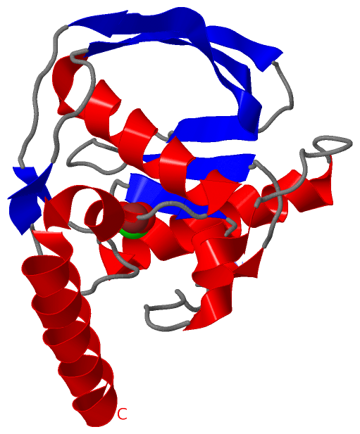
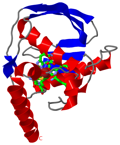
1dff PEPTIDE DEFORMYLASE
1dtf PEPTIDE DEFORMYLASE:THIORPHAN DOCKING MODEL 1
1g27 CRYSTAL STRUCTURE OF E.COLI POLYPEPTIDE DEFORMYLASECOMPLEXED WITH THE INHIBITOR BB- 3497
1g2a THE CRYSTAL STRUCTURE OF E.COLI PEPTIDE DEFORMYLASECOMPLEXED WITH ACTINONIN
1icj PDF PROTEIN IS CRYSTALLIZED AS NI2+ CONTAINING FORM,COCRYSTALLIZED WITH INHIBITOR POLYETHYLENE GLYCOL (PEG)
1lru CRYSTAL STRUCTURE OF E.COLI PEPTIDE DEFORMYLASE COMPLEXEDWITH ANTIBIOTIC ACTINONIN
2ai8 E.COLI POLYPEPTIDE DEFORMYLASE COMPLEXED WITH SB-485343
2def PEPTIDE DEFORMYLASE CATALYTIC CORE (RESIDUES 1 - 147), NMR, 20 STRUCTURES
2dtf PEPTIDE DEFORMYLASE:THIORPHAN DOCKING MODEL 1
2vhm STRUCTURE OF PDF BINDING HELIX IN COMPLEX WITH THE RIBOSOME
2w3t CHLORO COMPLEX OF THE NI-FORM OF E.COLI DEFORMYLASE