Show PDB file:   
         Plain Text   HTML   (compressed file size)
QuickSearch:   
by PDB,NDB,UniProt,PROSITE Code or Search Term(s)  
(-)Asymmetric Unit
(-)Asym. Unit - sites
(-)Biological Unit 1
(-)Biol. Unit 1 - sites
(-)Biological Unit 2
(-)Biological Unit 3
(-)Biological Unit 4
collapse expand < >
Image Asymmetric Unit
Asymmetric Unit  (Jmol Viewer)
Image Asym. Unit - sites
Asym. Unit - sites  (Jmol Viewer)
Image Biological Unit 1
Biological Unit 1  (Jmol Viewer)
Image Biol. Unit 1 - sites
Biol. Unit 1 - sites  (Jmol Viewer)
Image Biological Unit 2
Biological Unit 2  (Jmol Viewer)
Image Biological Unit 3
Biological Unit 3  (Jmol Viewer)
Image Biological Unit 4
Biological Unit 4  (Jmol Viewer)

(-) Description

Title :  CRYSTAL STRUCTURE OF BARLEY THIOREDOXIN H ISOFORM 1 CRYSTALLIZED USING AMMONIUM SULFATE AS PRECIPITANT
 
Authors :  K. Maeda, P. Hagglund, C. Finnie, B. Svensson, A. Henriksen
Date :  21 Jan 08  (Deposition) - 29 Apr 08  (Release) - 12 Jul 17  (Revision)
Method :  X-RAY DIFFRACTION
Resolution :  1.70
Chains :  Asym. Unit :  A,B,C,D
Biol. Unit 1:  A  (1x)
Biol. Unit 2:  B  (1x)
Biol. Unit 3:  C  (1x)
Biol. Unit 4:  D  (1x)
Keywords :  Oxidoreductase, Protein Disulfide Reductase, Thioredoxin-Fold (Keyword Search: [Gene Ontology, PubMed, Web (Google))
 
Reference :  K. Maeda, P. Hagglund, C. Finnie, B. Svensson, A. Henriksen
Crystal Structures Of Barley Thioredoxin H Isoforms Hvtrxh1 And Hvtrxh2 Reveal Features Involved In Protein Recognition And Possibly In Discriminating The Isoform Specificity.
Protein Sci. V. 17 1015 2008
PubMed-ID: 18424513  |  Reference-DOI: 10.1110/PS.083460308

(-) Compounds

Molecule 1 - THIOREDOXIN H ISOFORM 1.
    ChainsA, B, C, D
    EC Number1.8.1.9
    EngineeredYES
    Expression SystemESCHERICHIA COLI
    Expression System PlasmidPET11A
    Expression System StrainBL21(DE3)
    Expression System Taxid469008
    Expression System VariantROSETTA
    Organism CommonTWO-ROWED BARLEY
    Organism ScientificHORDEUM VULGARE VAR. DISTICHUM
    Organism Taxid112509
    SynonymTHIOREDOXIN
    VariantBARKE

 Structural Features

(-) Chains, Units

  1234
Asymmetric Unit ABCD
Biological Unit 1 (1x)A   
Biological Unit 2 (1x) B  
Biological Unit 3 (1x)  C 
Biological Unit 4 (1x)   D

Summary Information (see also Sequences/Alignments below)

(-) Ligands, Modified Residues, Ions  (1, 4)

Asymmetric Unit (1, 4)
No.NameCountTypeFull Name
1SO44Ligand/IonSULFATE ION
Biological Unit 1 (0, 0)
No.NameCountTypeFull Name
1SO4-1Ligand/IonSULFATE ION
Biological Unit 2 (1, 1)
No.NameCountTypeFull Name
1SO41Ligand/IonSULFATE ION
Biological Unit 3 (1, 1)
No.NameCountTypeFull Name
1SO41Ligand/IonSULFATE ION
Biological Unit 4 (1, 2)
No.NameCountTypeFull Name
1SO42Ligand/IonSULFATE ION

(-) Sites  (4, 4)

Asymmetric Unit (4, 4)
No.NameEvidenceResiduesDescription
1AC1SOFTWAREGLY D:100 , ARG D:101 , LYS D:102 , ASP D:103 , HOH D:2110BINDING SITE FOR RESIDUE SO4 D1116
2AC2SOFTWAREGLY C:100 , ARG C:101 , LYS C:102 , ASP C:103 , HOH C:2110BINDING SITE FOR RESIDUE SO4 C1119
3AC3SOFTWARELYS D:108BINDING SITE FOR RESIDUE SO4 D1117
4AC4SOFTWAREGLY B:100 , ARG B:101 , LYS B:102 , ASP B:103 , HOH B:2133 , PRO C:48BINDING SITE FOR RESIDUE SO4 B1116

(-) SS Bonds  (4, 4)

Asymmetric Unit
No.Residues
1A:40 -A:43
2B:40 -B:43
3C:40 -C:43
4D:40 -D:43

(-) Cis Peptide Bonds  (4, 6)

Asymmetric Unit
No.Residues
1Met A:82 -Pro A:83
2Met B:82 -Pro B:83
3Met C:82 -Pro C:83
4Met D:82 -Pro D:83

 Sequence-Structure Mapping

(-) SAPs(SNPs)/Variants  (0, 0)

(no "SAP(SNP)/Variant" information available for 2VM1)

(-) PROSITE Motifs  (0, 0)

(no "PROSITE Motif" information available for 2VM1)

(-) Exons   (0, 0)

(no "Exon" information available for 2VM1)

(-) Sequences/Alignments

Asymmetric Unit
   Reformat: Number of residues per line =  ('0' or empty: single-line sequence representation)
  Number of residues per labelling interval =   
  UniProt sequence: complete  aligned part    
   Show mapping: SCOP domains CATH domains Pfam domains Secondary structure (by author)
SAPs(SNPs) PROSITE motifs Exons
(details for a mapped element are shown in a popup box when the mouse pointer rests over it)
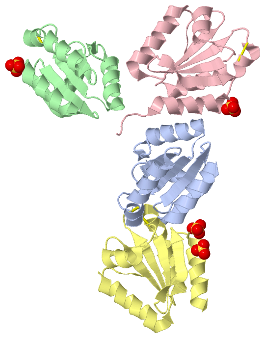
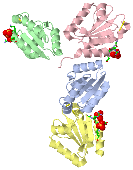
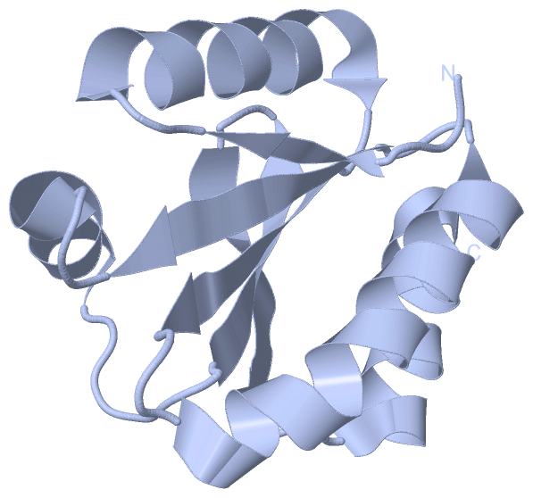
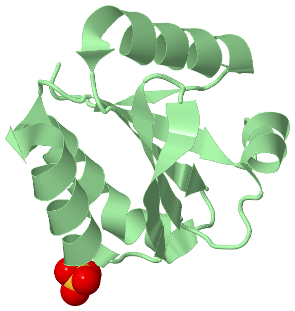
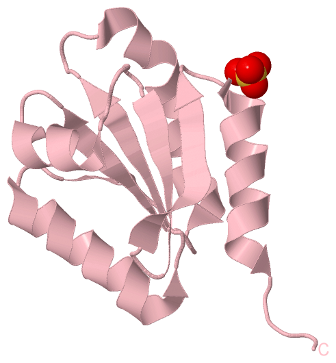
Chain A from PDB  Type:PROTEIN  Length:110
 aligned with Q7XZK3_HORVV | Q7XZK3 from UniProtKB/TrEMBL  Length:118

    Alignment length:110
                                    14        24        34        44        54        64        74        84        94       104       114
         Q7XZK3_HORVV     5 EGAVIACHTKQEFDTHMANGKDTGKLVIIDFTASWCGPCRVIAPVFAEYAKKFPGAIFLKVDVDELKDVAEAYNVEAMPTFLFIKDGEKVDSVVGGRKDDIHTKIVALMG 114
               SCOP domains d2vm1a_ A: automated matches                                                                                   SCOP domains
               CATH domains -------------------------------------------------------------------------------------------------------------- CATH domains
               Pfam domains -------------------------------------------------------------------------------------------------------------- Pfam domains
         Sec.struct. author ...eee..hhhhhhhhhhhhhhhh..eeeeee...hhhhhhhhhhhhhhhhhh...eeeeee...hhhhhhhh......eeeeee..eeeeeee..hhhhhhhhhhhhhh Sec.struct. author
                 SAPs(SNPs) -------------------------------------------------------------------------------------------------------------- SAPs(SNPs)
                    PROSITE -------------------------------------------------------------------------------------------------------------- PROSITE
                 Transcript -------------------------------------------------------------------------------------------------------------- Transcript
                 2vm1 A   5 EGAVIACHTKQEFDTHMANGKDTGKLVIIDFTASWCGPCRVIAPVFAEYAKKFPGAIFLKVDVDELKDVAEAYNVEAMPTFLFIKDGEKVDSVVGGRKDDIHTKIVALMG 114
                                    14        24        34        44        54        64        74        84        94       104       114

Chain B from PDB  Type:PROTEIN  Length:111
 aligned with Q7XZK3_HORVV | Q7XZK3 from UniProtKB/TrEMBL  Length:118

    Alignment length:111
                                    14        24        34        44        54        64        74        84        94       104       114 
         Q7XZK3_HORVV     5 EGAVIACHTKQEFDTHMANGKDTGKLVIIDFTASWCGPCRVIAPVFAEYAKKFPGAIFLKVDVDELKDVAEAYNVEAMPTFLFIKDGEKVDSVVGGRKDDIHTKIVALMGS 115
               SCOP domains d2vm1b_ B: automated matches                                                                                    SCOP domains
               CATH domains --------------------------------------------------------------------------------------------------------------- CATH domains
               Pfam domains --------------------------------------------------------------------------------------------------------------- Pfam domains
         Sec.struct. author ...eee..hhhhhhhhhhhhhhhh..eeeeee...hhhhhhhhhhhhhhhhhh...eeeeee...hhhhhhhh......eeeeee..eeeeeee..hhhhhhhhhhhhhh. Sec.struct. author
                 SAPs(SNPs) --------------------------------------------------------------------------------------------------------------- SAPs(SNPs)
                    PROSITE --------------------------------------------------------------------------------------------------------------- PROSITE
                 Transcript --------------------------------------------------------------------------------------------------------------- Transcript
                 2vm1 B   5 EGAVIACHTKQEFDTHMANGKDTGKLVIIDFTASWCGPCRVIAPVFAEYAKKFPGAIFLKVDVDELKDVAEAYNVEAMPTFLFIKDGEKVDSVVGGRKDDIHTKIVALMGS 115
                                    14        24        34        44        54        64        74        84        94       104       114 

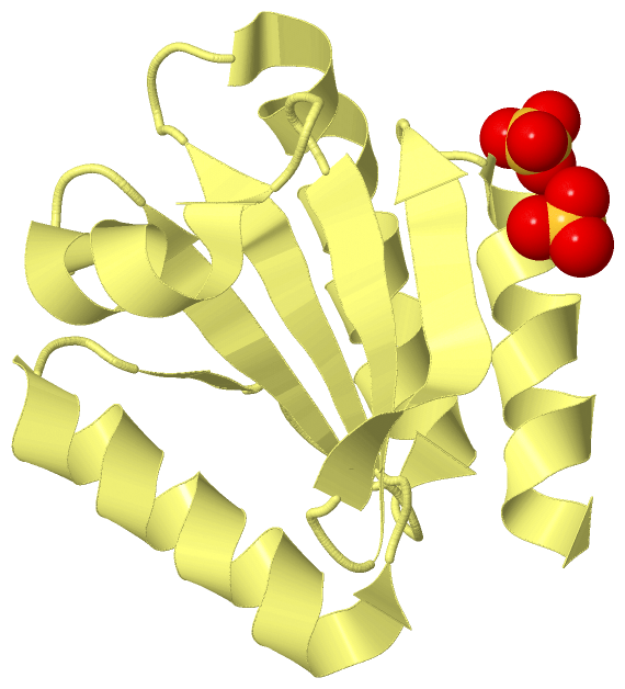
Chain C from PDB  Type:PROTEIN  Length:113
 aligned with Q7XZK3_HORVV | Q7XZK3 from UniProtKB/TrEMBL  Length:118

    Alignment length:113
                                    15        25        35        45        55        65        75        85        95       105       115   
         Q7XZK3_HORVV     6 GAVIACHTKQEFDTHMANGKDTGKLVIIDFTASWCGPCRVIAPVFAEYAKKFPGAIFLKVDVDELKDVAEAYNVEAMPTFLFIKDGEKVDSVVGGRKDDIHTKIVALMGSAST 118
               SCOP domains d2vm1c_ C: automated matches                                                                                      SCOP domains
               CATH domains ----------------------------------------------------------------------------------------------------------------- CATH domains
               Pfam domains ----------------------------------------------------------------------------------------------------------------- Pfam domains
         Sec.struct. author ...ee..hhhhhhhhhhhhhhhh..eeeeee...hhhhhhhhhhhhhhhhhh...eeeeee...hhhhhhhh......eeeeee..eeeeeee..hhhhhhhhhhhhhh.... Sec.struct. author
                 SAPs(SNPs) ----------------------------------------------------------------------------------------------------------------- SAPs(SNPs)
                    PROSITE ----------------------------------------------------------------------------------------------------------------- PROSITE
                 Transcript ----------------------------------------------------------------------------------------------------------------- Transcript
                 2vm1 C   6 GAVIACHTKQEFDTHMANGKDTGKLVIIDFTASWCGPCRVIAPVFAEYAKKFPGAIFLKVDVDELKDVAEAYNVEAMPTFLFIKDGEKVDSVVGGRKDDIHTKIVALMGSAST 118
                                    15        25        35        45        55        65        75        85        95       105       115   

Chain D from PDB  Type:PROTEIN  Length:110
 aligned with Q7XZK3_HORVV | Q7XZK3 from UniProtKB/TrEMBL  Length:118

    Alignment length:110
                                    15        25        35        45        55        65        75        85        95       105       115
         Q7XZK3_HORVV     6 GAVIACHTKQEFDTHMANGKDTGKLVIIDFTASWCGPCRVIAPVFAEYAKKFPGAIFLKVDVDELKDVAEAYNVEAMPTFLFIKDGEKVDSVVGGRKDDIHTKIVALMGS 115
               SCOP domains d2vm1d_ D: automated matches                                                                                   SCOP domains
               CATH domains -------------------------------------------------------------------------------------------------------------- CATH domains
               Pfam domains -------------------------------------------------------------------------------------------------------------- Pfam domains
         Sec.struct. author ...ee..hhhhhhhhhhhhhhhh..eeeeee...hhhhhhhhhhhhhhhhhh...eeeeee...hhhhhhhhh.....eeeeee..eeeeeee..hhhhhhhhhhhhhh. Sec.struct. author
                 SAPs(SNPs) -------------------------------------------------------------------------------------------------------------- SAPs(SNPs)
                    PROSITE -------------------------------------------------------------------------------------------------------------- PROSITE
                 Transcript -------------------------------------------------------------------------------------------------------------- Transcript
                 2vm1 D   6 GAVIACHTKQEFDTHMANGKDTGKLVIIDFTASWCGPCRVIAPVFAEYAKKFPGAIFLKVDVDELKDVAEAYNVEAMPTFLFIKDGEKVDSVVGGRKDDIHTKIVALMGS 115
                                    15        25        35        45        55        65        75        85        95       105       115

   Legend:   → Mismatch (orange background)
  - → Gap (green background, '-', border residues have a numbering label)
    → Modified Residue (blue background, lower-case, 'x' indicates undefined single-letter code, labelled with number + name)
  x → Chemical Group (purple background, 'x', labelled with number + name, e.g. ACE or NH2)
  extra numbering lines below/above indicate numbering irregularities and modified residue names etc., number ends below/above '|'

 Classification and Annotation

(-) SCOP Domains  (1, 4)

Asymmetric Unit

(-) CATH Domains  (0, 0)

(no "CATH Domain" information available for 2VM1)

(-) Pfam Domains  (0, 0)

(no "Pfam Domain" information available for 2VM1)

(-) Gene Ontology  (5, 5)

Asymmetric Unit(hide GO term definitions)
Chain A,B,C,D   (Q7XZK3_HORVV | Q7XZK3)
molecular function
    GO:0015035    protein disulfide oxidoreductase activity    Catalysis of the reaction: a protein with reduced sulfide groups = a protein with oxidized disulfide bonds.
biological process
    GO:0045454    cell redox homeostasis    Any process that maintains the redox environment of a cell or compartment within a cell.
    GO:0006662    glycerol ether metabolic process    The chemical reactions and pathways involving glycerol ethers, any anhydride formed between two organic hydroxy compounds, one of which is glycerol.
    GO:0055114    oxidation-reduction process    A metabolic process that results in the removal or addition of one or more electrons to or from a substance, with or without the concomitant removal or addition of a proton or protons.
cellular component
    GO:0005623    cell    The basic structural and functional unit of all organisms. Includes the plasma membrane and any external encapsulating structures such as the cell wall and cell envelope.

 Visualization

(-) Interactive Views

Asymmetric Unit
  Complete Structure
    Jena3D(integrated viewing of ligand, site, SAP, PROSITE, SCOP information)
    WebMol | AstexViewer[tm]@PDBe
(Java Applets, require no local installation except for Java; loading may be slow)
    STRAP
(Java WebStart application, automatic local installation, requires Java; full application with system access!)
    RasMol
(require local installation)
    Molscript (VRML)
(requires installation of a VRML viewer; select preferred view via VRML and generate a mono or stereo PDF format file)
 
  Ligands, Modified Residues, Ions
    SO4  [ RasMol | Jena3D ]  +environment [ RasMol | Jena3D ]
 
  Sites
    AC1  [ RasMol ]  +environment [ RasMol ]
    AC2  [ RasMol ]  +environment [ RasMol ]
    AC3  [ RasMol ]  +environment [ RasMol ]
    AC4  [ RasMol ]  +environment [ RasMol ]
 
  Cis Peptide Bonds
    Met A:82 - Pro A:83   [ RasMol ]  
    Met B:82 - Pro B:83   [ RasMol ]  
    Met C:82 - Pro C:83   [ RasMol ]  
    Met D:82 - Pro D:83   [ RasMol ]  
 
Biological Units
  Complete Structure
    Biological Unit 1  [ Jena3D ]
    Biological Unit 2  [ Jena3D ]
    Biological Unit 3  [ Jena3D ]
    Biological Unit 4  [ Jena3D ]

(-) Still Images

Jmol
  protein: cartoon or spacefill or dots and stick; nucleic acid: cartoon and stick; ligands: spacefill; active site: stick
Molscript
  protein, nucleic acid: cartoon; ligands: spacefill; active site: ball and stick

 Databases and Analysis Tools

(-) Databases

Access by PDB/NDB ID
  2vm1
    Family and Domain InformationProDom | SYSTERS
    General Structural InformationGlycoscienceDB | MMDB | NDB | OCA | PDB | PDBe | PDBj | PDBsum | PDBWiki | PQS | PROTEOPEDIA
    Orientation in MembranesOPM
    Protein SurfaceSURFACE
    Secondary StructureDSSP (structure derived) | HSSP (homology derived)
    Structural GenomicsGeneCensus
    Structural NeighboursCE | VAST
    Structure ClassificationCATH | Dali | SCOP
    Validation and Original DataBMRB Data View | BMRB Restraints Grid | EDS | PROCHECK | RECOORD | WHAT_CHECK
 
Access by UniProt ID/Accession number
  Q7XZK3_HORVV | Q7XZK3
    Comparative Protein Structure ModelsModBase
    Genomic InformationEnsembl
    Protein-protein InteractionDIP
    Sequence, Family and Domain InformationInterPro | Pfam | SMART | UniProtKB/TrEMBL
 
Access by Enzyme Classificator   (EC Number)
  1.8.1.9
    General Enzyme InformationBRENDA | EC-PDB | Enzyme | IntEnz
    PathwayKEGG | MetaCyc
 
Access by Disease Identifier   (MIM ID)
  (no 'MIM ID' available)
    Disease InformationOMIM
 
Access by GenAge ID
  (no 'GenAge ID' available)
    Age Related InformationGenAge

(-) Analysis Tools

Access by PDB/NDB ID
    Domain InformationXDom
    Interatomic Contacts of Structural UnitsCSU
    Ligand-protein ContactsLPC
    Protein CavitiescastP
    Sequence and Secondary StructurePDBCartoon
    Structure AlignmentSTRAP(Java WebStart application, automatic local installation, requires Java; full application with system access!)
    Structure and Sequence BrowserSTING
 
Access by UniProt ID/Accession number
  Q7XZK3_HORVV | Q7XZK3
    Protein Disorder PredictionDisEMBL | FoldIndex | GLOBPLOT (for more information see DisProt)

 Related Entries

(-) Entries Sharing at Least One Protein Chain (UniProt ID)

UniProtKB/TrEMBL
        Q7XZK3_HORVV | Q7XZK32vm2

(-) Related Entries Specified in the PDB File

2vm2 CRYSTAL STRUCTURE OF BARLEY THIOREDOXIN H ISOFORM 1 CRYSTALLIZED USING PEG AS PRECIPITANT