Show PDB file:   
         Plain Text   HTML   (compressed file size)
QuickSearch:   
by PDB,NDB,UniProt,PROSITE Code or Search Term(s)  
(-)Biol.Unit 1 - manually
(-)Asymmetric Unit
(-)Asym. Unit - sites
(-)Biological Unit 1
(-)Biol. Unit 1 - sites
collapse expand < >
Image Biol.Unit 1 - manually
Biol.Unit 1 - manually  (Jmol Viewer)
Image Asymmetric Unit
Asymmetric Unit  (Jmol Viewer)
Image Asym. Unit - sites
Asym. Unit - sites  (Jmol Viewer)
Image Biological Unit 1
Biological Unit 1  (Jmol Viewer)
Image Biol. Unit 1 - sites
Biol. Unit 1 - sites  (Jmol Viewer)

(-) Description

Title :  HELIX BUNDLE QUATERNARY STRUCTURE FROM ALPHA/BETA-PEPTIDE FOLDAMERS: GCN4-PLI WITH BETA-RESIDUES AT B AND F HEPTAD POSITIONS.
 
Authors :  W. S. Horne, J. L. Price, J. L. Keck, S. H. Gellman
Date :  20 Feb 07  (Deposition) - 27 Mar 07  (Release) - 13 Jul 11  (Revision)
Method :  X-RAY DIFFRACTION
Resolution :  2.00
Chains :  Asym. Unit :  A
Biol. Unit 1:  A  (4x)
Keywords :  Helix Bundle, Foldamer, Unknown Function (Keyword Search: [Gene Ontology, PubMed, Web (Google))
 
Reference :  W. S. Horne, J. L. Price, J. L. Keck, S. H. Gellman
Helix Bundle Quaternary Structure From Alpha/Beta-Peptide Foldamers.
J. Am. Chem. Soc. V. 129 4178 2007
PubMed-ID: 17362016  |  Reference-DOI: 10.1021/JA070396F

(-) Compounds

Molecule 1 - HYBRID ALPHA/BETA PEPTIDE BASED ON THE GCN4-PLI SEQUENCE
    ChainsA
    EngineeredYES
    Other DetailsSYNTHETIC PEPTIDE
    SyntheticYES

 Structural Features

(-) Chains, Units

  1
Asymmetric Unit A
Biological Unit 1 (4x)A

Summary Information (see also Sequences/Alignments below)

(-) Ligands, Modified Residues, Ions  (10, 15)

Asymmetric Unit (10, 15)
No.NameCountTypeFull Name
1ACE1Mod. Amino AcidACETYL GROUP
2B3A1Mod. Amino Acid(3S)-3-AMINOBUTANOIC ACID
3B3D1Mod. Amino Acid3-AMINOPENTANEDIOIC ACID
4B3E1Mod. Amino Acid(3S)-3-AMINOHEXANEDIOIC ACID
5B3K2Mod. Amino Acid(3S)-3,7-DIAMINOHEPTANOIC ACID
6B3S1Mod. Amino Acid(3R)-3-AMINO-4-HYDROXYBUTANOIC ACID
7B3X1Mod. Amino Acid(3S)-3,5-DIAMINO-5-OXOPENTANOIC ACID
8B3Y1Mod. Amino Acid(3S)-3-AMINO-4-(4-HYDROXYPHENYL)BUTANOIC ACID
9BAL1Mod. Amino AcidBETA-ALANINE
10FMT5Ligand/IonFORMIC ACID
Biological Unit 1 (10, 60)
No.NameCountTypeFull Name
1ACE4Mod. Amino AcidACETYL GROUP
2B3A4Mod. Amino Acid(3S)-3-AMINOBUTANOIC ACID
3B3D4Mod. Amino Acid3-AMINOPENTANEDIOIC ACID
4B3E4Mod. Amino Acid(3S)-3-AMINOHEXANEDIOIC ACID
5B3K8Mod. Amino Acid(3S)-3,7-DIAMINOHEPTANOIC ACID
6B3S4Mod. Amino Acid(3R)-3-AMINO-4-HYDROXYBUTANOIC ACID
7B3X4Mod. Amino Acid(3S)-3,5-DIAMINO-5-OXOPENTANOIC ACID
8B3Y4Mod. Amino Acid(3S)-3-AMINO-4-(4-HYDROXYPHENYL)BUTANOIC ACID
9BAL4Mod. Amino AcidBETA-ALANINE
10FMT20Ligand/IonFORMIC ACID

(-) Sites  (5, 5)

Asymmetric Unit (5, 5)
No.NameEvidenceResiduesDescription
1AC1SOFTWAREB3S A:14 , B3Y A:17 , ARG A:25 , B3K A:28 , LEU A:29BINDING SITE FOR RESIDUE FMT A 34
2AC2SOFTWAREB3X A:21 , B3A A:24 , B3K A:28 , HOH A:39BINDING SITE FOR RESIDUE FMT A 35
3AC3SOFTWAREB3K A:3 , ARG A:33BINDING SITE FOR RESIDUE FMT A 36
4AC4SOFTWAREARG A:1 , LEU A:13 , B3S A:14 , GLU A:32BINDING SITE FOR RESIDUE FMT A 37
5AC5SOFTWAREARG A:25 , LYS A:27BINDING SITE FOR RESIDUE FMT A 38

(-) SS Bonds  (0, 0)

(no "SS Bond" information available for 2OXK)

(-) Cis Peptide Bonds  (0, 0)

(no "Cis Peptide Bond" information available for 2OXK)

 Sequence-Structure Mapping

(-) SAPs(SNPs)/Variants  (0, 0)

(no "SAP(SNP)/Variant" information available for 2OXK)

(-) PROSITE Motifs  (0, 0)

(no "PROSITE Motif" information available for 2OXK)

(-) Exons   (0, 0)

(no "Exon" information available for 2OXK)

(-) Sequences/Alignments

Asymmetric Unit
   Reformat: Number of residues per line =  ('0' or empty: single-line sequence representation)
  Number of residues per labelling interval =   
  UniProt sequence: complete  aligned part    
   Show mapping: SCOP domains CATH domains Pfam domains Secondary structure (by author)
SAPs(SNPs) PROSITE motifs Exons
(details for a mapped element are shown in a popup box when the mouse pointer rests over it)
Chain A from PDB  Type:PROTEIN  Length:34
                                                                 
               SCOP domains ---------------------------------- SCOP domains
               CATH domains ---------------------------------- CATH domains
               Pfam domains ---------------------------------- Pfam domains
         Sec.struct. author .hhhhhhhhhhhhhhhhhhhhhhhhhhhhhh... Sec.struct. author
                 SAPs(SNPs) ---------------------------------- SAPs(SNPs)
                    PROSITE ---------------------------------- PROSITE
                 Transcript ---------------------------------- Transcript
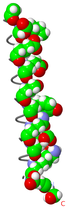
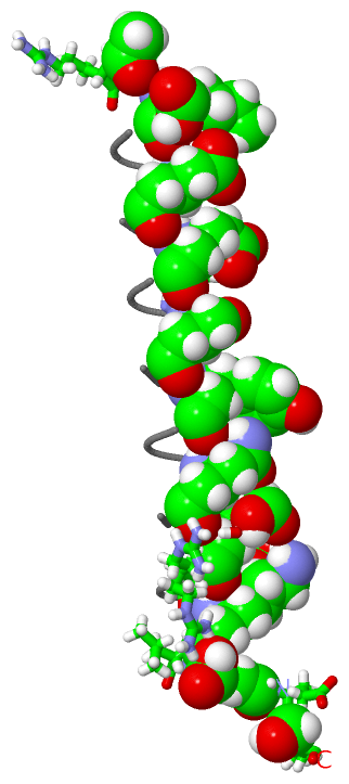
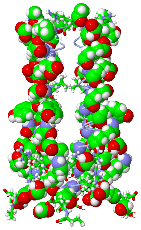
                  2oxk A  0 xRMkQIEdKLeEILsKLyHIEnELaRIKkLLaER 33
                            |  |   | 9|   |  |19 |  |   29 |  
                            |  |   |  |   |  |   |  |   |  |  
                            0-ACE  |  |   |  |   |  |   |  |  
                               3-B3K  |   |  |   |  |   |  |  
                                   7-B3D  |  |   |  |   |  |  
                                     10-B3E  |   |  |   |  |  
                                         14-B3S  |  |   |  |  
                                            17-B3Y  |   |  |  
                                                21-B3X  |  |  
                                                   24-B3A  |  

   Legend:   → Mismatch (orange background)
  - → Gap (green background, '-', border residues have a numbering label)
    → Modified Residue (blue background, lower-case, 'x' indicates undefined single-letter code, labelled with number + name)
  x → Chemical Group (purple background, 'x', labelled with number + name, e.g. ACE or NH2)
  extra numbering lines below/above indicate numbering irregularities and modified residue names etc., number ends below/above '|'

 Classification and Annotation

(-) SCOP Domains  (0, 0)

(no "SCOP Domain" information available for 2OXK)

(-) CATH Domains  (0, 0)

(no "CATH Domain" information available for 2OXK)

(-) Pfam Domains  (0, 0)

(no "Pfam Domain" information available for 2OXK)

(-) Gene Ontology  (0, 0)

Asymmetric Unit(hide GO term definitions)
    (no "Gene Ontology" information available for 2OXK)

 Visualization

(-) Interactive Views

Asymmetric Unit
  Complete Structure
    Jena3D(integrated viewing of ligand, site, SAP, PROSITE, SCOP information)
    WebMol | AstexViewer[tm]@PDBe
(Java Applets, require no local installation except for Java; loading may be slow)
    STRAP
(Java WebStart application, automatic local installation, requires Java; full application with system access!)
    RasMol
(require local installation)
    Molscript (VRML)
(requires installation of a VRML viewer; select preferred view via VRML and generate a mono or stereo PDF format file)
 
  Ligands, Modified Residues, Ions
    ACE  [ RasMol | Jena3D ]  +environment [ RasMol | Jena3D ]
    B3A  [ RasMol | Jena3D ]  +environment [ RasMol | Jena3D ]
    B3D  [ RasMol | Jena3D ]  +environment [ RasMol | Jena3D ]
    B3E  [ RasMol | Jena3D ]  +environment [ RasMol | Jena3D ]
    B3K  [ RasMol | Jena3D ]  +environment [ RasMol | Jena3D ]
    B3S  [ RasMol | Jena3D ]  +environment [ RasMol | Jena3D ]
    B3X  [ RasMol | Jena3D ]  +environment [ RasMol | Jena3D ]
    B3Y  [ RasMol | Jena3D ]  +environment [ RasMol | Jena3D ]
    BAL  [ RasMol | Jena3D ]  +environment [ RasMol | Jena3D ]
    FMT  [ RasMol | Jena3D ]  +environment [ RasMol | Jena3D ]
 
  Sites
    AC1  [ RasMol ]  +environment [ RasMol ]
    AC2  [ RasMol ]  +environment [ RasMol ]
    AC3  [ RasMol ]  +environment [ RasMol ]
    AC4  [ RasMol ]  +environment [ RasMol ]
    AC5  [ RasMol ]  +environment [ RasMol ]
 
  Cis Peptide Bonds
(no "Cis Peptide Bonds" information available for 2oxk)
 
Biological Unit
  Complete Structure
    Biological Unit 1  [ Jena3D ]

(-) Still Images

Jmol
  protein: cartoon or spacefill or dots and stick; nucleic acid: cartoon and stick; ligands: spacefill; active site: stick
Molscript
  protein, nucleic acid: cartoon; ligands: spacefill; active site: ball and stick

 Databases and Analysis Tools

(-) Databases

Access by PDB/NDB ID
  2oxk
    Family and Domain InformationProDom | SYSTERS
    General Structural InformationGlycoscienceDB | MMDB | NDB | OCA | PDB | PDBe | PDBj | PDBsum | PDBWiki | PQS | PROTEOPEDIA
    Orientation in MembranesOPM
    Protein SurfaceSURFACE
    Secondary StructureDSSP (structure derived) | HSSP (homology derived)
    Structural GenomicsGeneCensus
    Structural NeighboursCE | VAST
    Structure ClassificationCATH | Dali | SCOP
    Validation and Original DataBMRB Data View | BMRB Restraints Grid | EDS | PROCHECK | RECOORD | WHAT_CHECK
 
Access by UniProt ID/Accession number
  (no 'UniProt ID/Accession number' available) |
    Comparative Protein Structure ModelsModBase
    Genomic InformationEnsembl
    Protein-protein InteractionDIP
    Sequence, Family and Domain InformationInterPro | Pfam | SMART | UniProt
 
Access by Enzyme Classificator   (EC Number)
  (no 'Enzyme Classificator' available)
    General Enzyme InformationBRENDA | EC-PDB | Enzyme | IntEnz
    PathwayKEGG | MetaCyc
 
Access by Disease Identifier   (MIM ID)
  (no 'MIM ID' available)
    Disease InformationOMIM
 
Access by GenAge ID
  (no 'GenAge ID' available)
    Age Related InformationGenAge

(-) Analysis Tools

Access by PDB/NDB ID
    Domain InformationXDom
    Interatomic Contacts of Structural UnitsCSU
    Ligand-protein ContactsLPC
    Protein CavitiescastP
    Sequence and Secondary StructurePDBCartoon
    Structure AlignmentSTRAP(Java WebStart application, automatic local installation, requires Java; full application with system access!)
    Structure and Sequence BrowserSTING
 
Access by UniProt ID/Accession number
  (no 'UniProt ID/Accession number' available) |
    Protein Disorder PredictionDisEMBL | FoldIndex | GLOBPLOT

 Related Entries

(-) Entries Sharing at Least One Protein Chain (UniProt ID)

(no "Entries Sharing at Least One Protein Chain" available for 2OXK)

(-) Related Entries Specified in the PDB File

2oxj