Show PDB file:   
         Plain Text   HTML   (compressed file size)
QuickSearch:   
by PDB,NDB,UniProt,PROSITE Code or Search Term(s)  
(-)NMR Structure - model 1
(-)NMR Structure - all models
collapse expand < >
Image NMR Structure - model 1
NMR Structure - model 1  (Jmol Viewer)
Image NMR Structure - all models
NMR Structure - all models  (Jmol Viewer)

(-) Description

Title :  3D STRUCTURE AND BACKBONE DYNAMICS OF SPE B
 
Authors :  W. Chuang, C. Wang, H. Houng, C. Chen, P. Wang
Date :  26 Jul 07  (Deposition) - 26 Aug 08  (Release) - 21 Jul 09  (Revision)
Method :  SOLUTION NMR
Resolution :  NOT APPLICABLE
Chains :  NMR Structure  :  A  (20x)
Keywords :  Dynamics, Loop Movement, Nmr, Streptopain, Structure, Hydrolase, Protease, Secreted, Thiol Protease, Toxin, Virulence, Zymogen (Keyword Search: [Gene Ontology, PubMed, Web (Google))
 
Reference :  C. C. Wang, H. C. Houng, C. L. Chen, P. J. Wang, C. F. Kuo, Y. S. Lin, J. J. Wu, M. T. Lin, C. C. Liu, W. Huang, W. J. Chuang
Solution Structure And Backbone Dynamics Of Streptopain: Insight Into Diverse Substrate Specificity.
J. Biol. Chem. V. 284 10957 2009
PubMed-ID: 19237546  |  Reference-DOI: 10.1074/JBC.M807624200
(for further references see the PDB file header)

(-) Compounds

Molecule 1 - STREPTOPAIN
    ChainsA
    EC Number3.4.22.10
    EngineeredYES
    Expression SystemESCHERICHIA COLI
    Expression System StrainBL21(DE3)PLYSS
    Expression System Taxid562
    Expression System VectorPET21A
    GeneSPEB
    MutationYES
    Organism ScientificSTREPTOCOCCUS PYOGENES SEROTYPE M1
    Organism Taxid301447
    StrainA-20 (M1T1)
    SynonymSTREPTOCOCCAL CYSTEINE PROTEINASE, STREPTOCOCCUS PEPTIDASE A, SPP, EXOTOXIN TYPE B, SPE B

 Structural Features

(-) Chains, Units

  
NMR Structure (20x)

Summary Information (see also Sequences/Alignments below)

(-) Ligands, Modified Residues, Ions  (0, 0)

(no "Ligand,Modified Residues,Ions" information available for 2JTC)

(-) Sites  (0, 0)

(no "Site" information available for 2JTC)

(-) SS Bonds  (0, 0)

(no "SS Bond" information available for 2JTC)

(-) Cis Peptide Bonds  (0, 0)

(no "Cis Peptide Bond" information available for 2JTC)

 Sequence-Structure Mapping

(-) SAPs(SNPs)/Variants  (2, 2)

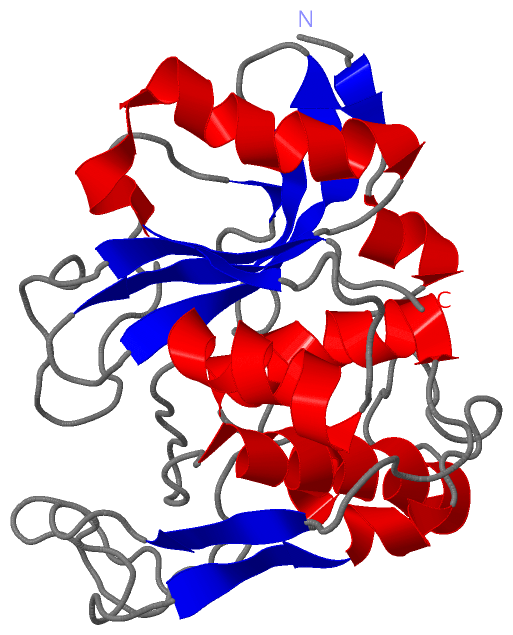
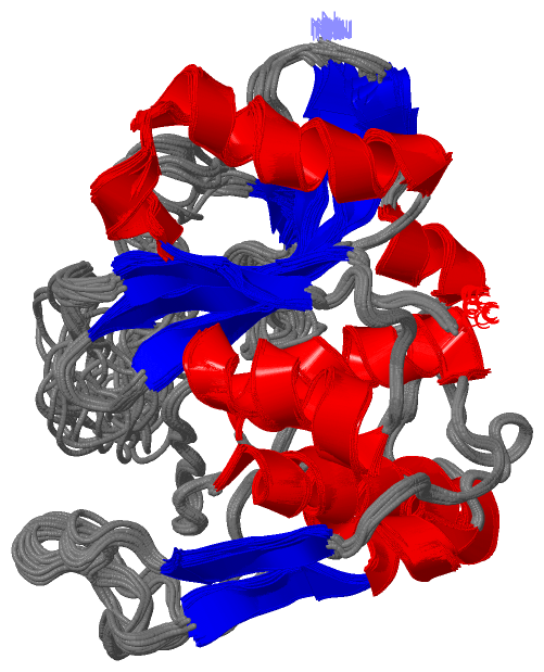
NMR Structure (2, 2)
  dbSNPPDB
No.SourceVariant IDVariantUniProt IDStatusIDChainVariant
1UniProtVAR_SPEB_STRP1_002 *V193LSPEB_STRP1  ---  ---AV48L
2UniProtVAR_SPEB_STRP1_003 *G308SSPEB_STRP1  ---  ---AG163S
   * ID not provided by source

  SNP/SAP Summary Statistics (UniProtKB/Swiss-Prot)

(-) PROSITE Motifs  (0, 0)

(no "PROSITE Motif" information available for 2JTC)

(-) Exons   (0, 0)

(no "Exon" information available for 2JTC)

(-) Sequences/Alignments

NMR Structure
   Reformat: Number of residues per line =  ('0' or empty: single-line sequence representation)
  Number of residues per labelling interval =   
  UniProt sequence: complete  aligned part    
   Show mapping: SCOP domains CATH domains Pfam domains Secondary structure (by author)
SAPs(SNPs) PROSITE motifs Exons
(details for a mapped element are shown in a popup box when the mouse pointer rests over it)
Chain A from PDB  Type:PROTEIN  Length:253
 aligned with SPEB_STRP1 | P0C0J1 from UniProtKB/Swiss-Prot  Length:398

    Alignment length:253
                                   155       165       175       185       195       205       215       225       235       245       255       265       275       285       295       305       315       325       335       345       355       365       375       385       395   
           SPEB_STRP1   146 QPVVKSLLDSKGIHYNQGNPYNLLTPVIEKVKPGEQSFVGQHAATGCVATATAQIMKYHNYPNKGLKDYTYTLSSNNPYFNHPKNLFAAISTRQYNWNNILPTYSGRESNVQKMAISELMADVGISVDMDYGPSSGSAGSSRVQRALKENFGYNQSVHQINRGDFSKQDWEAQIDKELSQNQPVYYQGVGKVGGHAFVIDGADGRNFYHVNWGWGGVSDGFFRLDALNPSALGTGGGAGGFNGYQSAVVGIKP 398
               SCOP domains ------------------------------------------------------------------------------------------------------------------------------------------------------------------------------------------------------------------------------------------------------------- SCOP domains
               CATH domains ------------------------------------------------------------------------------------------------------------------------------------------------------------------------------------------------------------------------------------------------------------- CATH domains
               Pfam domains Inhibitor_I---Peptidase_C10-2jtcA01 A:15-223                                                                                                                                                                                   ------------------------------ Pfam domains
         Sec.struct. author ..ee.hhhhhh.......hhhhh......................hhhhhhhhhhhhhhh........eeee............eeeehhhhh...............hhhhhhhhhhhhhhhhhhh...........hhhhhhhhhhhh.......eeehhhhhhhhhhhhhhhhhhhh...eeeeee....eeeeeeeeee....eeee........ee.......................eeeee.... Sec.struct. author
                 SAPs(SNPs) -----------------------------------------------L------------------------------------------------------------------------------------------------------------------S------------------------------------------------------------------------------------------ SAPs(SNPs)
                    PROSITE ------------------------------------------------------------------------------------------------------------------------------------------------------------------------------------------------------------------------------------------------------------- PROSITE
                 Transcript ------------------------------------------------------------------------------------------------------------------------------------------------------------------------------------------------------------------------------------------------------------- Transcript
                 2jtc A   1 QPVVKSLLDSKGIHYNQGNPYNLLTPVIEKVKPGEQSFVGQHAATGSVATATAQIMKYHNYPNKGLKDYTYTLSSNNPYFNHPKNLFAAISTRQYNWNNILPTYSGRESNVQKMAISELMADVGISVDMDYGPSSGSAGSSRVQRALKENFGYNQSVHQINRGDFSKQDWEAQIDKELSQNQPVYYQGVGKVGGHAFVIDGADGRNFYHVNWGWGGVSDGFFRLDALNPSALGTGGGAGGFNGYQSAVVGIKP 253
                                    10        20        30        40        50        60        70        80        90       100       110       120       130       140       150       160       170       180       190       200       210       220       230       240       250   

   Legend:   → Mismatch (orange background)
  - → Gap (green background, '-', border residues have a numbering label)
    → Modified Residue (blue background, lower-case, 'x' indicates undefined single-letter code, labelled with number + name)
  x → Chemical Group (purple background, 'x', labelled with number + name, e.g. ACE or NH2)
  extra numbering lines below/above indicate numbering irregularities and modified residue names etc., number ends below/above '|'

 Classification and Annotation

(-) SCOP Domains  (0, 0)

(no "SCOP Domain" information available for 2JTC)

(-) CATH Domains  (0, 0)

(no "CATH Domain" information available for 2JTC)

(-) Pfam Domains  (2, 2)

NMR Structure

(-) Gene Ontology  (6, 6)

NMR Structure(hide GO term definitions)
Chain A   (SPEB_STRP1 | P0C0J1)
molecular function
    GO:0008234    cysteine-type peptidase activity    Catalysis of the hydrolysis of peptide bonds in a polypeptide chain by a mechanism in which the sulfhydryl group of a cysteine residue at the active center acts as a nucleophile.
    GO:0016787    hydrolase activity    Catalysis of the hydrolysis of various bonds, e.g. C-O, C-N, C-C, phosphoric anhydride bonds, etc. Hydrolase is the systematic name for any enzyme of EC class 3.
    GO:0008233    peptidase activity    Catalysis of the hydrolysis of a peptide bond. A peptide bond is a covalent bond formed when the carbon atom from the carboxyl group of one amino acid shares electrons with the nitrogen atom from the amino group of a second amino acid.
biological process
    GO:0009405    pathogenesis    The set of specific processes that generate the ability of an organism to induce an abnormal, generally detrimental state in another organism.
    GO:0006508    proteolysis    The hydrolysis of proteins into smaller polypeptides and/or amino acids by cleavage of their peptide bonds.
cellular component
    GO:0005576    extracellular region    The space external to the outermost structure of a cell. For cells without external protective or external encapsulating structures this refers to space outside of the plasma membrane. This term covers the host cell environment outside an intracellular parasite.

 Visualization

(-) Interactive Views

NMR Structure
  Complete Structure
    Jena3D(integrated viewing of ligand, site, SAP, PROSITE, SCOP information)
    WebMol | AstexViewer[tm]@PDBe
(Java Applets, require no local installation except for Java; loading may be slow)
    STRAP
(Java WebStart application, automatic local installation, requires Java; full application with system access!)
    RasMol
(require local installation)
    Molscript (VRML)
(requires installation of a VRML viewer; select preferred view via VRML and generate a mono or stereo PDF format file)
 
  Ligands, Modified Residues, Ions
(no "Ligands, Modified Residues, Ions" information available for 2jtc)
 
  Sites
(no "Sites" information available for 2jtc)
 
  Cis Peptide Bonds
(no "Cis Peptide Bonds" information available for 2jtc)
 

(-) Still Images

Jmol
  protein: cartoon or spacefill or dots and stick; nucleic acid: cartoon and stick; ligands: spacefill; active site: stick
Molscript
  protein, nucleic acid: cartoon; ligands: spacefill; active site: ball and stick

 Databases and Analysis Tools

(-) Databases

Access by PDB/NDB ID
  2jtc
    Family and Domain InformationProDom | SYSTERS
    General Structural InformationGlycoscienceDB | MMDB | NDB | OCA | PDB | PDBe | PDBj | PDBsum | PDBWiki | PQS | PROTEOPEDIA
    Orientation in MembranesOPM
    Protein SurfaceSURFACE
    Secondary StructureDSSP (structure derived) | HSSP (homology derived)
    Structural GenomicsGeneCensus
    Structural NeighboursCE | VAST
    Structure ClassificationCATH | Dali | SCOP
    Validation and Original DataBMRB Data View | BMRB Restraints Grid | EDS | PROCHECK | RECOORD | WHAT_CHECK
 
Access by UniProt ID/Accession number
  SPEB_STRP1 | P0C0J1
    Comparative Protein Structure ModelsModBase
    Genomic InformationEnsembl
    Protein-protein InteractionDIP
    Sequence, Family and Domain InformationInterPro | Pfam | SMART | UniProtKB/SwissProt
 
Access by Enzyme Classificator   (EC Number)
  3.4.22.10
    General Enzyme InformationBRENDA | EC-PDB | Enzyme | IntEnz
    PathwayKEGG | MetaCyc
 
Access by Disease Identifier   (MIM ID)
  (no 'MIM ID' available)
    Disease InformationOMIM
 
Access by GenAge ID
  (no 'GenAge ID' available)
    Age Related InformationGenAge

(-) Analysis Tools

Access by PDB/NDB ID
    Domain InformationXDom
    Interatomic Contacts of Structural UnitsCSU
    Ligand-protein ContactsLPC
    Protein CavitiescastP
    Sequence and Secondary StructurePDBCartoon
    Structure AlignmentSTRAP(Java WebStart application, automatic local installation, requires Java; full application with system access!)
    Structure and Sequence BrowserSTING
 
Access by UniProt ID/Accession number
  SPEB_STRP1 | P0C0J1
    Protein Disorder PredictionDisEMBL | FoldIndex | GLOBPLOT (for more information see DisProt)

 Related Entries

(-) Entries Sharing at Least One Protein Chain (UniProt ID)

(no "Entries Sharing at Least One Protein Chain" available for 2JTC)

(-) Related Entries Specified in the PDB File

5547