|
|
|
|
 Description
|
|
Compounds
|
||||||||||||||||||||||||||||||||||||||||||||||||||
Chains, Units
Summary Information (see also Sequences/Alignments below) |
Ligands, Modified Residues, Ions (5, 45)
Asymmetric Unit (5, 45)
|
Sites (7, 7)
Asymmetric Unit (7, 7)
|
SS Bonds (0, 0)| (no "SS Bond" information available for 2G32) |
Cis Peptide Bonds (0, 0)| (no "Cis Peptide Bond" information available for 2G32) |
SAPs(SNPs)/Variants (0, 0)| (no "SAP(SNP)/Variant" information available for 2G32) |
PROSITE Motifs (0, 0)| (no "PROSITE Motif" information available for 2G32) |
Exons (0, 0)| (no "Exon" information available for 2G32) |
Sequences/Alignments
Asymmetric Unit
Chain K from PDB  Type:OTHER  Length:8

                  2g32 K 79 xxxxxxxx 86
                            ||||||||
                            ||||||||
                           79-0C||||
                            80-0U|||
                             81-0G||
                              82-0G|
                               83-0G
                                84-0C
                                 85-0G
                                  86-0G
Chain L from PDB  Type:OTHER  Length:8

                  2g32 L 90 xxxxxxxx 97
                            ||||||||
                            ||||||||
                           90-0C||||
                            91-0C|||
                             92-0G||
                              93-0C|
                               94-0C
                                95-0U
                                 96-0G
                                  97-0G
Chain M from PDB  Type:OTHER  Length:8

                  2g32 M 79 xxxxxxxx 86
                            ||||||||
                            ||||||||
                           79-0C||||
                            80-0U|||
                             81-0G||
                              82-0G|
                               83-0G
                                84-0C
                                 85-0G
                                  86-0G
Chain N from PDB  Type:OTHER  Length:8

                  2g32 N 90 xxxxxxxx 97
                            ||||||||
                            ||||||||
                           90-0C||||
                            91-0C|||
                             92-0G||
                              93-0C|
                               94-0C
                                95-0U
                                 96-0G
                                  97-0G
|
||||||||||||||||||||
SCOP Domains (0, 0)| (no "SCOP Domain" information available for 2G32) |
CATH Domains (0, 0)| (no "CATH Domain" information available for 2G32) |
Pfam Domains (0, 0)| (no "Pfam Domain" information available for 2G32) |
Gene Ontology (0, 0)|
Asymmetric Unit(hide GO term definitions)
(no "Gene Ontology" information available for 2G32)
|
Interactive Views
|
|||||||||||||||||||||||||||||||||||||||||||||||||||||||||||||||||||||||||||||||||||||||||||||||||||||||||||||||||||||||||||||||||||||||||||||||||||||||||||||||||||||||||||||||||||||||||||||||||||||||||||||||||||
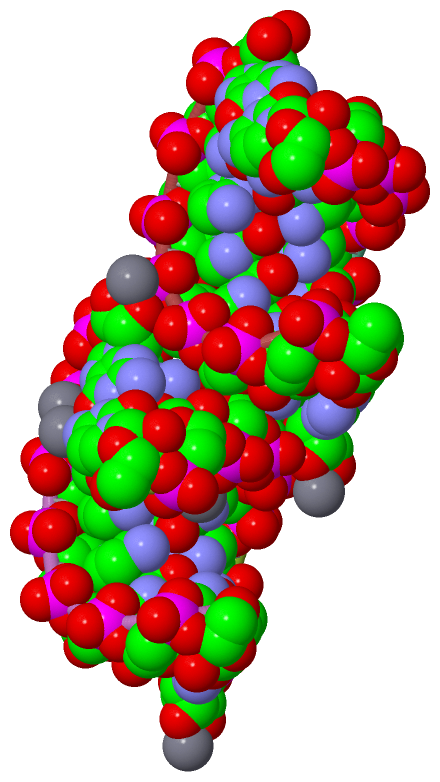
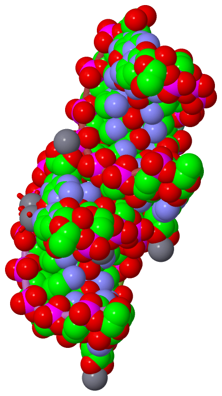
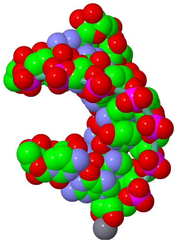
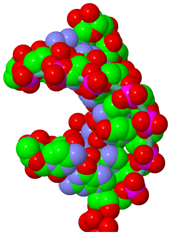
Still Images
|
||||||||||||||||
Databases
|
||||||||||||||||||||||||||||||||||||||||||||||||||||||||||||||||||||||||||||||||||||||||||||||||||||||||||||||||||||||||||||||||||||||||||||||||||||||||||||||||
Analysis Tools
|
|||||||||||||||||||||||||||||||||||||||||||||||||||||||||||||
Entries Sharing at Least One Protein Chain (UniProt ID)
Related Entries Specified in the PDB File
|
|