Asymmetric/Biological Unit(hide GO term definitions)
Chain A ( Q672W7_HELPX | Q672W7)
| molecular function |
|---|
| | GO:0016787 | | hydrolase activity | | Catalysis of the hydrolysis of various bonds, e.g. C-O, C-N, C-C, phosphoric anhydride bonds, etc. Hydrolase is the systematic name for any enzyme of EC class 3. |
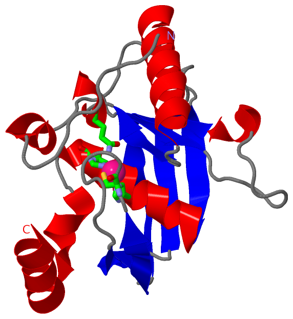
| | GO:0005506 | | iron ion binding | | Interacting selectively and non-covalently with iron (Fe) ions. |
| | GO:0046872 | | metal ion binding | | Interacting selectively and non-covalently with any metal ion. |
| | GO:0042586 | | peptide deformylase activity | | Catalysis of the reaction: formyl-L-methionyl peptide + H2O = formate + methionyl peptide. |
| biological process |
|---|
| | GO:0006412 | | translation | | The cellular metabolic process in which a protein is formed, using the sequence of a mature mRNA or circRNA molecule to specify the sequence of amino acids in a polypeptide chain. Translation is mediated by the ribosome, and begins with the formation of a ternary complex between aminoacylated initiator methionine tRNA, GTP, and initiation factor 2, which subsequently associates with the small subunit of the ribosome and an mRNA or circRNA. Translation ends with the release of a polypeptide chain from the ribosome. |
Chain A ( Q2L8P7_HELPX | Q2L8P7)
| molecular function |
|---|
| | GO:0016787 | | hydrolase activity | | Catalysis of the hydrolysis of various bonds, e.g. C-O, C-N, C-C, phosphoric anhydride bonds, etc. Hydrolase is the systematic name for any enzyme of EC class 3. |
| | GO:0005506 | | iron ion binding | | Interacting selectively and non-covalently with iron (Fe) ions. |
| | GO:0046872 | | metal ion binding | | Interacting selectively and non-covalently with any metal ion. |
| | GO:0042586 | | peptide deformylase activity | | Catalysis of the reaction: formyl-L-methionyl peptide + H2O = formate + methionyl peptide. |
| biological process |
|---|
| | GO:0006412 | | translation | | The cellular metabolic process in which a protein is formed, using the sequence of a mature mRNA or circRNA molecule to specify the sequence of amino acids in a polypeptide chain. Translation is mediated by the ribosome, and begins with the formation of a ternary complex between aminoacylated initiator methionine tRNA, GTP, and initiation factor 2, which subsequently associates with the small subunit of the ribosome and an mRNA or circRNA. Translation ends with the release of a polypeptide chain from the ribosome. |
|
 Description