|
|
|
|
 Description
|
|
Compounds
|
||||||||||||||||||||||||||||||||||||||||||||||||
Chains, Units
Summary Information (see also Sequences/Alignments below) |
Ligands, Modified Residues, Ions (1, 2)
NMR Structure (1, 2)
|
Sites (2, 2)
NMR Structure (2, 2)
|
SS Bonds (0, 0)| (no "SS Bond" information available for 2ECN) |
Cis Peptide Bonds (0, 0)| (no "Cis Peptide Bond" information available for 2ECN) |
SAPs(SNPs)/Variants (0, 0)| (no "SAP(SNP)/Variant" information available for 2ECN) |
PROSITE Motifs (2, 2)
NMR Structure (2, 2)
|
||||||||||||||||||||||||||||||||
Exons (3, 3)
NMR Structure (3, 3)
|
||||||||||||||||||||||||||||||||||||||||||||||||||||||||||||||||||||||||||||||||||||||||||||||||
Sequences/Alignments
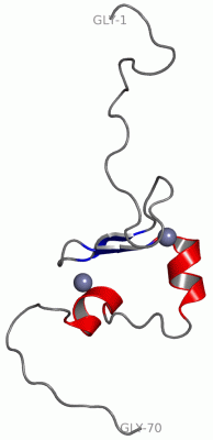
NMR StructureChain A from PDB Type:PROTEIN Length:70 aligned with RN141_HUMAN | Q8WVD5 from UniProtKB/Swiss-Prot Length:230 Alignment length:93 128 138 148 158 168 178 188 198 208 RN141_HUMAN 119 AQSSTSEEPDENSSSVTSCQASLWMGRVKQLTDEEECCICMDGRADLILPCAHSFCQKCIDKWSDRHRNCPICRLQMTGANESWVVSDAPTED 211 SCOP domains --------------------------------------------------------------------------------------------- SCOP domains CATH domains --------------------------------------------------------------------------------------------- CATH domains Pfam domains --------------------------------------------------------------------------------------------- Pfam domains SAPs(SNPs) --------------------------------------------------------------------------------------------- SAPs(SNPs) PROSITE (1) ------------------------------------ZF_RING_2 PDB: A:18-55 ------------------- PROSITE (1) PROSITE (2) --------------------------------------------------ZF_RING_1 --------------------------------- PROSITE (2) Transcript 1 (1) Exon 1.6 PDB: A:1-8 (gaps)-----------------------------------Exon 1.8b PDB: A:44-70 (gaps) Transcript 1 (1) Transcript 1 (2) --------------------------Exon 1.7a PDB: A:8-44 ------------------------------ Transcript 1 (2) 2ecn A 1 GSSGSS-------------------GRVKQLTDEEECCICMDGRADLILPCAHSFCQKCIDKWSDRHRNCPICRLQMTGANE----SSGPSSG 70 | - - | 11 21 31 41 51 61 | | 67 6 7 63 64
|
||||||||||||||||||||
SCOP Domains (0, 0)| (no "SCOP Domain" information available for 2ECN) |
CATH Domains (0, 0)| (no "CATH Domain" information available for 2ECN) |
Pfam Domains (0, 0)| (no "Pfam Domain" information available for 2ECN) |
Gene Ontology (7, 7)|
NMR Structure(hide GO term definitions) Chain A (RN141_HUMAN | Q8WVD5)
|
||||||||||||||||||||||||||||||||||||||||||||||||||||||||||||
Interactive Views
|
|||||||||||||||||||||||||||||||||||||||||||||||||||||||||||||||||||||||||||||||||||||||||||||||||||||||||||||||||||||||||||||
Still Images
|
||||||||||||||||
Databases
|
||||||||||||||||||||||||||||||||||||||||||||||||||||||||||||||||||||||||||||||||||||||||||||||||||||||||||||||||||||||||||||||||||||||||||||||||||||||||||||||||
Analysis Tools
|
|||||||||||||||||||||||||||||||||||||||||||||||||||||||||||||
Entries Sharing at Least One Protein Chain (UniProt ID)
Related Entries Specified in the PDB File
|
|