|
|
|
|
 Description
|
|
Compounds
|
||||||||||||||||||||||||||||||||||||||||||||
Chains, Units
Summary Information (see also Sequences/Alignments below) |
Ligands, Modified Residues, Ions (1, 1)
Asymmetric/Biological Unit (1, 1)
|
Sites (1, 1)
Asymmetric Unit (1, 1)
|
SS Bonds (1, 1)
Asymmetric/Biological Unit
|
||||||||
Cis Peptide Bonds (1, 1)
Asymmetric/Biological Unit
|
||||||||
SAPs(SNPs)/Variants (0, 0)| (no "SAP(SNP)/Variant" information available for 2CBM) |
PROSITE Motifs (0, 0)| (no "PROSITE Motif" information available for 2CBM) |
Exons (0, 0)| (no "Exon" information available for 2CBM) |
Sequences/Alignments
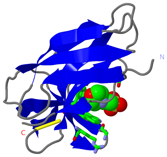
Asymmetric/Biological UnitChain A from PDB Type:PROTEIN Length:112 aligned with NCZS_STRCZ | P0A3R9 from UniProtKB/Swiss-Prot Length:147 Alignment length:112 44 54 64 74 84 94 104 114 124 134 144 NCZS_STRCZ 35 AAPTATVTPSSGLSDGTVVKVAGAGLQAGTAYDVGQCAWVDTGVLACNPADFSSVTADANGSASTSLTVRRSFEGFLFDGTRWGTVDCTTAACQVGLSDAAGNGPEGVAISF 146 SCOP domains d2cbma_ A: automated matches SCOP domains CATH domains 2cbmA00 A:1-112 [code=2.60.40.230, no name defined] CATH domains Pfam domains ---------------------------------------------------------------------------------------------------------------- Pfam domains SAPs(SNPs) ---------------------------------------------------------------------------------------------------------------- SAPs(SNPs) PROSITE ---------------------------------------------------------------------------------------------------------------- PROSITE Transcript ---------------------------------------------------------------------------------------------------------------- Transcript 2cbm A 1 AAPTATVTPSSGLSDGTVVKVAGAGLQAGTAYWVAQWARVDTGVWAYNPADNSSVTADANGSASTSLTVRRSFEGFLFDGTRWGTVDCTTAACQVGLSDAAGNGPEGVAISF 112 10 20 30 40 50 60 70 80 90 100 110
|
||||||||||||||||||||
SCOP Domains (1, 1)
Asymmetric/Biological Unit
 |
CATH Domains (1, 1)
Asymmetric/Biological Unit
|
Pfam Domains (0, 0)| (no "Pfam Domain" information available for 2CBM) |
Gene Ontology (3, 3)|
Asymmetric/Biological Unit(hide GO term definitions) Chain A (NCZS_STRCZ | P0A3R9)
|
||||||||||||||||||||||||||||||
Interactive Views
|
|||||||||||||||||||||||||||||||||||||||||||||||||||||||||||||||||||||||||||||||||||||||||||||||||||||||||||||||||||||||
Still Images
|
||||||||||||||||
Databases
|
||||||||||||||||||||||||||||||||||||||||||||||||||||||||||||||||||||||||||||||||||||||||||||||||||||||||||||||||||||||||||||||||||||||||||||||||||||||||||||||||
Analysis Tools
|
|||||||||||||||||||||||||||||||||||||||||||||||||||||||||||||
Entries Sharing at Least One Protein Chain (UniProt ID)
Related Entries Specified in the PDB File
|
|