|
|
|
|
 Description
|
|
Compounds
|
||||||||||||||||||||||||||||||||||||||||||||||||||||||||
Chains, Units
Summary Information (see also Sequences/Alignments below) |
Ligands, Modified Residues, Ions (0, 0)| (no "Ligand,Modified Residues,Ions" information available for 2B1U) |
Sites (0, 0)| (no "Site" information available for 2B1U) |
SS Bonds (0, 0)| (no "SS Bond" information available for 2B1U) |
Cis Peptide Bonds (0, 0)| (no "Cis Peptide Bond" information available for 2B1U) |
SAPs(SNPs)/Variants (0, 0)| (no "SAP(SNP)/Variant" information available for 2B1U) |
PROSITE Motifs (2, 4)
NMR Structure (2, 4)
|
||||||||||||||||||||||||||||||||
Exons (1, 1)
NMR Structure (1, 1)
|
||||||||||||||||||||||||||||||||||||
Sequences/Alignments
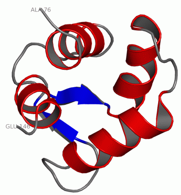
NMR StructureChain A from PDB Type:PROTEIN Length:71 aligned with CALL5_HUMAN | Q9NZT1 from UniProtKB/Swiss-Prot Length:146 Alignment length:71 85 95 105 115 125 135 145 CALL5_HUMAN 76 ARAGLEDLQVAFRAFDQDGDGHITVDELRRAMAGLGQPLPQEELDAMIREADVDQDGRVNYEEFARMLAQE 146 SCOP domains d2b1ua_ A: automated matches SCOP domains CATH domains ----------------------------------------------------------------------- CATH domains Pfam domains ----------------------------------------------------------------------- Pfam domains SAPs(SNPs) ----------------------------------------------------------------------- SAPs(SNPs) PROSITE (1) --EF_HAND_2 PDB: A:78-113 EF_HAND_2 PDB: A:114-146 PROSITE (1) PROSITE (2) ---------------EF_HAND_1 -----------------------EF_HAND_1 ------- PROSITE (2) Transcript 1 Exon 1.1 PDB: A:76-146 UniProt: 1-183 [INCOMPLETE] Transcript 1 2b1u A 76 ARAGLEDLQVAFRAFDQDGDGHITVDELRRAMAGLGQPLPQEELDAMIREADVDQDGRVNYEEFARMLAQE 146 85 95 105 115 125 135 145
|
||||||||||||||||||||
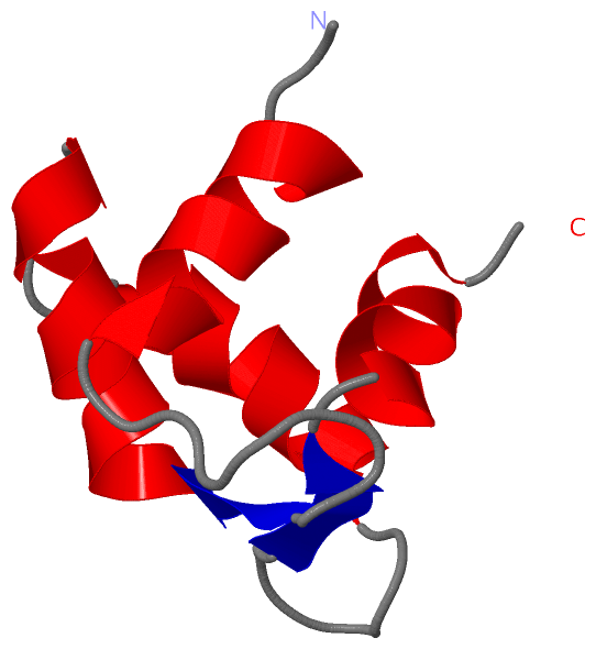
SCOP Domains (1, 1)
NMR Structure
 |
CATH Domains (0, 0)| (no "CATH Domain" information available for 2B1U) |
Pfam Domains (0, 0)| (no "Pfam Domain" information available for 2B1U) |
Gene Ontology (5, 5)|
NMR Structure(hide GO term definitions) Chain A (CALL5_HUMAN | Q9NZT1)
|
||||||||||||||||||||||||||||||||||||||||||||||||
Interactive Views
|
||||||||||||||||||||||||||||||||||||||||||||||||||||||||||||||||||||||||||||||||||||||||||||||||||||||||||||||||||||
Still Images
|
||||||||||||||||
Databases
|
||||||||||||||||||||||||||||||||||||||||||||||||||||||||||||||||||||||||||||||||||||||||||||||||||||||||||||||||||||||||||||||||||||||||||||||||||||||||||||||||
Analysis Tools
|
|||||||||||||||||||||||||||||||||||||||||||||||||||||||||||||
Entries Sharing at Least One Protein Chain (UniProt ID)
Related Entries Specified in the PDB File
|
|