Show PDB file:   
         Plain Text   HTML   (compressed file size)
QuickSearch:   
by PDB,NDB,UniProt,PROSITE Code or Search Term(s)  
(-)Asym./Biol. Unit
(-)Asym./Biol. Unit - sites
collapse expand < >
Image Asym./Biol. Unit
Asym./Biol. Unit  (Jmol Viewer)
Image Asym./Biol. Unit - sites
Asym./Biol. Unit - sites  (Jmol Viewer)

(-) Description

Title :  CRYSTAL STRUCTURE OF THE LIGANDED CHITIN OLIGASACCHARIDE BINDING PROTEIN
 
Authors :  S. Xu, X. Li, R. Roseman, A. M. Stock
Date :  28 May 05  (Deposition) - 06 Jun 06  (Release) - 24 Feb 09  (Revision)
Method :  X-RAY DIFFRACTION
Resolution :  2.20
Chains :  Asym./Biol. Unit :  A
Keywords :  Alpha Helix/Beta Sheet, Sugar Binding Protein, Signaling Protein (Keyword Search: [Gene Ontology, PubMed, Web (Google))
 
Reference :  S. Xu, X. Li, R. Roseman, A. M. Stock
Crystal Structure Of The Liganded And Unliganded Periplasmic Chitin Oligosaccharide Binding Protein
To Be Published
PubMed: search
(for further references see the PDB file header)

(-) Compounds

Molecule 1 - CHITIN OLIGOSACCHARIDE BINDING PROTEIN
    ChainsA
    EngineeredYES
    Expression SystemESCHERICHIA COLI BL21(DE3)
    Expression System PlasmidPMAL-P2
    Expression System StrainBL21(DE3)
    Expression System Taxid469008
    Expression System Vector TypePLASMID
    FragmentRESIDUES 28-556
    Organism ScientificVIBRIO CHOLERAE
    Organism Taxid666

 Structural Features

(-) Chains, Units

  1
Asymmetric/Biological Unit A

Summary Information (see also Sequences/Alignments below)
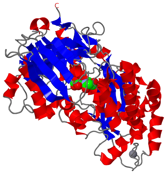
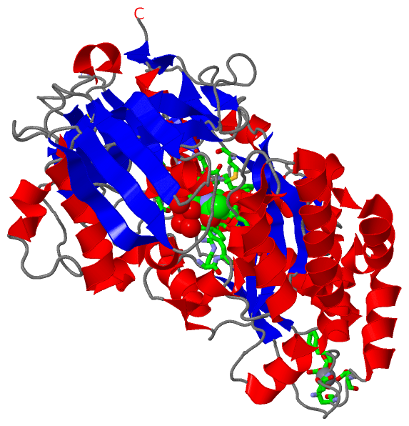
(-) Ligands, Modified Residues, Ions  (2, 2)

Asymmetric/Biological Unit (2, 2)
No.NameCountTypeFull Name
1CBS1Ligand/IonDI(N-ACETYL-D-GLUCOSAMINE)
2MN1Ligand/IonMANGANESE (II) ION

(-) Sites  (2, 2)

Asymmetric Unit (2, 2)
No.NameEvidenceResiduesDescription
1AC1SOFTWAREVAL A:7 , PRO A:8 , ASP A:9 , PHE A:10 , ASN A:203 , SER A:219 , SER A:220 , PHE A:221 , TRP A:362 , ASP A:364 , PHE A:365 , ASN A:408 , TYR A:409 , ARG A:435 , PHE A:436 , MET A:488 , TRP A:512 , HOH A:905 , HOH A:957 , HOH A:988 , HOH A:1034 , HOH A:1118 , HOH A:1509BINDING SITE FOR RESIDUE CBS A 901
2AC2SOFTWAREASP A:337 , ASN A:339 , ASP A:341 , PHE A:343 , GLU A:345BINDING SITE FOR RESIDUE MN A 801

(-) SS Bonds  (1, 1)

Asymmetric/Biological Unit
No.Residues
1A:179 -A:194

(-) Cis Peptide Bonds  (0, 0)

(no "Cis Peptide Bond" information available for 1ZU0)

 Sequence-Structure Mapping

(-) SAPs(SNPs)/Variants  (0, 0)

(no "SAP(SNP)/Variant" information available for 1ZU0)

(-) PROSITE Motifs  (0, 0)

(no "PROSITE Motif" information available for 1ZU0)

(-) Exons   (0, 0)

(no "Exon" information available for 1ZU0)

(-) Sequences/Alignments

Asymmetric/Biological Unit
   Reformat: Number of residues per line =  ('0' or empty: single-line sequence representation)
  Number of residues per labelling interval =   
  UniProt sequence: complete  aligned part    
   Show mapping: SCOP domains CATH domains Pfam domains Secondary structure (by author)
SAPs(SNPs) PROSITE motifs Exons
(details for a mapped element are shown in a popup box when the mouse pointer rests over it)
Chain A from PDB  Type:PROTEIN  Length:529
 aligned with Q9KUA3_VIBCH | Q9KUA3 from UniProtKB/TrEMBL  Length:556

    Alignment length:529
                                    37        47        57        67        77        87        97       107       117       127       137       147       157       167       177       187       197       207       217       227       237       247       257       267       277       287       297       307       317       327       337       347       357       367       377       387       397       407       417       427       437       447       457       467       477       487       497       507       517       527       537       547         
         Q9KUA3_VIBCH    28 RSELTIVPDFYPTMVRNFNPYLATNLRTTTDFIYEPLVVFNEMKGNTPVFRLAESYKMADDLMSVTFDIRKGVKWSDGEAFTADDVVYSFGLLKAKPELDQRGINKWVTSVEKVDEYKVRFRLSEANSNVPYEISLIPIVAEHVWKDVKDPTTFTNENPVGTGPFTVIDTFTPQLYIQCRNPNYWDAANLEVDCLRVPQIANNDQLLGKIVNSELDWTSSFVPDIDRTYAAANPNHHYWYPAAGTQAFMVNFKNPDPAKKEALDNVDFRRAFSMALDRQTIIDIAFYGSGTVNDFASGLGYAFEAWSDEATHKKYKGFNTYDVEGSKKLLAKAGFKDVNGDGFVETPSGKSFELLIQSPNGWTDFNNTVQLAVEQLQEVGIKAKARTPEFAVYNQAMLEGTYDVAYTNYFHGADPFTYWNSGYNSALQSGDGMPRFAMHYFTDKKLDGLLDSFYKTADKNEQLAIAHGIQKIIAENQVTIPVMSGAWMYQYNTTRFTGWWSEENPKGRPSVWAGIPERLLHVLDLKPVK 556
               SCOP domains ------------------------------------------------------------------------------------------------------------------------------------------------------------------------------------------------------------------------------------------------------------------------------------------------------------------------------------------------------------------------------------------------------------------------------------------------------------------------------------------------------------------------------------------------- SCOP domains
               CATH domains ------------------------------------------------------------------------------------------------------------------------------------------------------------------------------------------------------------------------------------------------------------------------------------------------------------------------------------------------------------------------------------------------------------------------------------------------------------------------------------------------------------------------------------------------- CATH domains
               Pfam domains ----------------------------------------------SBP_bac_5-1zu0A01 A:47-431                                                                                                                                                                                                                                                                                                                                                                       -------------------------------------------------------------------------------------------------- Pfam domains
         Sec.struct. author ..eeeee..............................eeee.....eeee..eeeeee.....eeeeee............hhhhhhhhhhhhhhhhhhh..hhhhheeeeeeee..eeeeee.....hhhhhhhh....hhhhhh..................eeeeeeee..eeeeee.....hhhhh...eeeee...hhhhhhhhhhh....ee.....hhhhhh......eee...eeeeeeeee.....hhhhhhhhhhhhhhhhhhhhhhhhhhhhhhh....ee.......hhhhhhhhhhhhhhhhhhhhh.hhhhhhhhhhhh....................eeee....hhhhhhhhhhhhhhhhhh...eeee..hhhhhhhhhhh....eeeee......hhhhhhhhhhhhhhh.............hhhhhhhhhhhhhh.hhhhhhhhhhhhhhhhhhhh..eeeeeee...eee...ee..................hhhhhhhh..ee.. Sec.struct. author
                 SAPs(SNPs) ------------------------------------------------------------------------------------------------------------------------------------------------------------------------------------------------------------------------------------------------------------------------------------------------------------------------------------------------------------------------------------------------------------------------------------------------------------------------------------------------------------------------------------------------- SAPs(SNPs)
                    PROSITE ------------------------------------------------------------------------------------------------------------------------------------------------------------------------------------------------------------------------------------------------------------------------------------------------------------------------------------------------------------------------------------------------------------------------------------------------------------------------------------------------------------------------------------------------- PROSITE
                 Transcript ------------------------------------------------------------------------------------------------------------------------------------------------------------------------------------------------------------------------------------------------------------------------------------------------------------------------------------------------------------------------------------------------------------------------------------------------------------------------------------------------------------------------------------------------- Transcript
                 1zu0 A   1 RSELTIVPDFYPTMVRNFNPYLATNLRTTTDFIYEPLVVFNEMKGNTPVFRLAESYKMADDLMSVTFDIRKGVKWSDGEAFTADDVVYSFGLLKAKPELDQRGINKWVTSVEKVDEYKVRFRLSEANSNVPYEISLIPIVAEHVWKDVKDPTTFTNENPVGTGPFTVIDTFTPQLYIQCRNPNYWDAANLEVDCLRVPQIANNDQLLGKIVNSELDWTSSFVPDIDRTYAAANPNHHYWYPAAGTQAFMVNFKNPDPAKKEALDNVDFRRAFSMALDRQTIIDIAFYGSGTVNDFASGLGYAFEAWSDEATHKKYKGFNTYDVEGSKKLLAKAGFKDVNGDGFVETPSGKSFELLIQSPNGWTDFNNTVQLAVEQLQEVGIKAKARTPEFAVYNQAMLEGTYDVAYTNYFHGADPFTYWNSGYNSALQSGDGMPRFAMHYFTDKKLDGLLDSFYKTADKNEQLAIAHGIQKIIAENQVTIPVMSGAWMYQYNTTRFTGWWSEENPKGRPSVWAGIPERLLHVLDLKPVK 529
                                    10        20        30        40        50        60        70        80        90       100       110       120       130       140       150       160       170       180       190       200       210       220       230       240       250       260       270       280       290       300       310       320       330       340       350       360       370       380       390       400       410       420       430       440       450       460       470       480       490       500       510       520         

   Legend:   → Mismatch (orange background)
  - → Gap (green background, '-', border residues have a numbering label)
    → Modified Residue (blue background, lower-case, 'x' indicates undefined single-letter code, labelled with number + name)
  x → Chemical Group (purple background, 'x', labelled with number + name, e.g. ACE or NH2)
  extra numbering lines below/above indicate numbering irregularities and modified residue names etc., number ends below/above '|'

 Classification and Annotation

(-) SCOP Domains  (0, 0)

(no "SCOP Domain" information available for 1ZU0)

(-) CATH Domains  (0, 0)

(no "CATH Domain" information available for 1ZU0)

(-) Pfam Domains  (1, 1)

Asymmetric/Biological Unit
(-)
Clan: PBP (391)

(-) Gene Ontology  (7, 7)

Asymmetric/Biological Unit(hide GO term definitions)
Chain A   (Q9KUA3_VIBCH | Q9KUA3)
molecular function
    GO:0042626    ATPase activity, coupled to transmembrane movement of substances    Catalysis of the reaction: ATP + H2O = ADP + phosphate, to directly drive the active transport of a substance across a membrane.
    GO:0046872    metal ion binding    Interacting selectively and non-covalently with any metal ion.
    GO:0015197    peptide transporter activity    Enables the directed movement of peptides, compounds of two or more amino acids where the alpha carboxyl group of one is bound to the alpha amino group of another, into, out of or within a cell, or between cells.
biological process
    GO:0015833    peptide transport    The directed movement of peptides, compounds of two or more amino acids where the alpha carboxyl group of one is bound to the alpha amino group of another, into, out of or within a cell, or between cells, by means of some agent such as a transporter or pore.
    GO:0055085    transmembrane transport    The process in which a solute is transported across a lipid bilayer, from one side of a membrane to the other
cellular component
    GO:0043190    ATP-binding cassette (ABC) transporter complex    A complex for the transport of metabolites into and out of the cell, typically comprised of four domains; two membrane-associated domains and two ATP-binding domains at the intracellular face of the membrane, that form a central pore through the plasma membrane. Each of the four core domains may be encoded as a separate polypeptide or the domains can be fused in any one of a number of ways into multidomain polypeptides. In Bacteria and Archaebacteria, ABC transporters also include substrate binding proteins to bind substrate external to the cytoplasm and deliver it to the transporter.
    GO:0030288    outer membrane-bounded periplasmic space    The region between the inner (cytoplasmic or plasma) membrane and outer membrane of organisms with two membranes such as Gram negative bacteria. These periplasmic spaces are relatively thick and contain a thin peptidoglycan layer (PGL), also referred to as a thin cell wall.

 Visualization

(-) Interactive Views

Asymmetric/Biological Unit
  Complete Structure
    Jena3D(integrated viewing of ligand, site, SAP, PROSITE, SCOP information)
    WebMol | AstexViewer[tm]@PDBe
(Java Applets, require no local installation except for Java; loading may be slow)
    STRAP
(Java WebStart application, automatic local installation, requires Java; full application with system access!)
    RasMol
(require local installation)
    Molscript (VRML)
(requires installation of a VRML viewer; select preferred view via VRML and generate a mono or stereo PDF format file)
 
  Ligands, Modified Residues, Ions
    CBS  [ RasMol | Jena3D ]  +environment [ RasMol | Jena3D ]
    MN  [ RasMol | Jena3D ]  +environment [ RasMol | Jena3D ]
 
  Sites
    AC1  [ RasMol ]  +environment [ RasMol ]
    AC2  [ RasMol ]  +environment [ RasMol ]
 
  Cis Peptide Bonds
(no "Cis Peptide Bonds" information available for 1zu0)
 

(-) Still Images

Jmol
  protein: cartoon or spacefill or dots and stick; nucleic acid: cartoon and stick; ligands: spacefill; active site: stick
Molscript
  protein, nucleic acid: cartoon; ligands: spacefill; active site: ball and stick

 Databases and Analysis Tools

(-) Databases

Access by PDB/NDB ID
  1zu0
    Family and Domain InformationProDom | SYSTERS
    General Structural InformationGlycoscienceDB | MMDB | NDB | OCA | PDB | PDBe | PDBj | PDBsum | PDBWiki | PQS | PROTEOPEDIA
    Orientation in MembranesOPM
    Protein SurfaceSURFACE
    Secondary StructureDSSP (structure derived) | HSSP (homology derived)
    Structural GenomicsGeneCensus
    Structural NeighboursCE | VAST
    Structure ClassificationCATH | Dali | SCOP
    Validation and Original DataBMRB Data View | BMRB Restraints Grid | EDS | PROCHECK | RECOORD | WHAT_CHECK
 
Access by UniProt ID/Accession number
  Q9KUA3_VIBCH | Q9KUA3
    Comparative Protein Structure ModelsModBase
    Genomic InformationEnsembl
    Protein-protein InteractionDIP
    Sequence, Family and Domain InformationInterPro | Pfam | SMART | UniProtKB/TrEMBL
 
Access by Enzyme Classificator   (EC Number)
  (no 'Enzyme Classificator' available)
    General Enzyme InformationBRENDA | EC-PDB | Enzyme | IntEnz
    PathwayKEGG | MetaCyc
 
Access by Disease Identifier   (MIM ID)
  (no 'MIM ID' available)
    Disease InformationOMIM
 
Access by GenAge ID
  (no 'GenAge ID' available)
    Age Related InformationGenAge

(-) Analysis Tools

Access by PDB/NDB ID
    Domain InformationXDom
    Interatomic Contacts of Structural UnitsCSU
    Ligand-protein ContactsLPC
    Protein CavitiescastP
    Sequence and Secondary StructurePDBCartoon
    Structure AlignmentSTRAP(Java WebStart application, automatic local installation, requires Java; full application with system access!)
    Structure and Sequence BrowserSTING
 
Access by UniProt ID/Accession number
  Q9KUA3_VIBCH | Q9KUA3
    Protein Disorder PredictionDisEMBL | FoldIndex | GLOBPLOT (for more information see DisProt)

 Related Entries

(-) Entries Sharing at Least One Protein Chain (UniProt ID)

UniProtKB/TrEMBL
        Q9KUA3_VIBCH | Q9KUA31zty 4gf8 4gfr

(-) Related Entries Specified in the PDB File

1zty THE SAME PROTEIN WITHOUT LIGAND