|
|
|
|
 Description
|
|
Compounds
|
||||||||||||||||||||
Chains, Units
Summary Information (see also Sequences/Alignments below) |
Ligands, Modified Residues, Ions (1, 1)
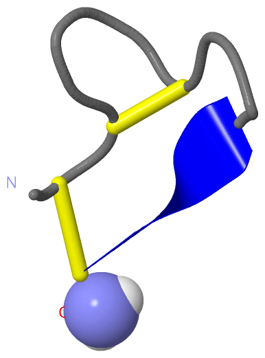
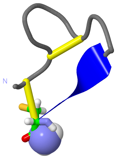
NMR Structure (1, 1)
|
Sites (1, 1)
NMR Structure (1, 1)
|
SS Bonds (2, 2)
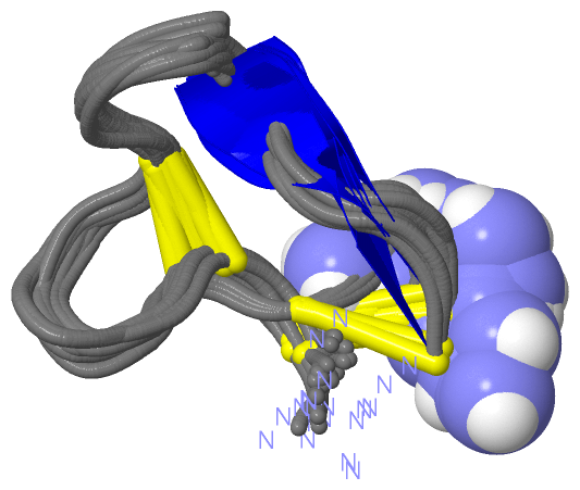
NMR Structure
|
||||||||||||
Cis Peptide Bonds (1, 24)
NMR Structure
|
||||||||||
SAPs(SNPs)/Variants (0, 0)| (no "SAP(SNP)/Variant" information available for 1XGB) |
PROSITE Motifs (1, 1)
NMR Structure (1, 1)
|
||||||||||||||||||||||||
Exons (0, 0)| (no "Exon" information available for 1XGB) |
Sequences/Alignments
NMR StructureChain A from PDB Type:PROTEIN Length:14 aligned with CA1A_CONGE | P01519 from UniProtKB/Swiss-Prot Length:15 Alignment length:14 10 CA1A_CONGE 1 ECCNPACGRHYSCG 14 SCOP domains d1xgba_ A: SCOP domains CATH domains -------------- CATH domains Pfam domains -------------- Pfam domains SAPs(SNPs) -------------- SAPs(SNPs) PROSITE -ALPHA_CONOTO- PROSITE Transcript -------------- Transcript 1xgb A 1 ECCNPACGRHYSCx 14 10 | 14-NH2
|
||||||||||||||||||||
SCOP Domains (1, 1)
NMR Structure
 |
CATH Domains (0, 0)| (no "CATH Domain" information available for 1XGB) |
Pfam Domains (0, 0)| (no "Pfam Domain" information available for 1XGB) |
Gene Ontology (2, 2)|
NMR Structure(hide GO term definitions) Chain A (CA1A_CONGE | P01519)
|
||||||||||||||||||
Interactive Views
|
|||||||||||||||||||||||||||||||||||||||||||||||||||||||||||||||||||||||||||||||||||||||||||||||||||||||||||||||||||||||
Still Images
|
||||||||||||||||
Databases
|
||||||||||||||||||||||||||||||||||||||||||||||||||||||||||||||||||||||||||||||||||||||||||||||||||||||||||||||||||||||||||||||||||||||||||||||||||||||||||||||||
Analysis Tools
|
|||||||||||||||||||||||||||||||||||||||||||||||||||||||||||||
Entries Sharing at Least One Protein Chain (UniProt ID)
Related Entries Specified in the PDB File
|
|