Show PDB file:   
         Plain Text   HTML   (compressed file size)
QuickSearch:   
by PDB,NDB,UniProt,PROSITE Code or Search Term(s)  
(-)Biol.Unit 1 - manually
(-)Asymmetric Unit
(-)Asym. Unit - sites
(-)Biological Unit 1
(-)Biol. Unit 1 - sites
collapse expand < >
Image Biol.Unit 1 - manually
Biol.Unit 1 - manually  (Jmol Viewer)
Image Asymmetric Unit
Asymmetric Unit  (Jmol Viewer)
Image Asym. Unit - sites
Asym. Unit - sites  (Jmol Viewer)
Image Biological Unit 1
Biological Unit 1  (Jmol Viewer)
Image Biol. Unit 1 - sites
Biol. Unit 1 - sites  (Jmol Viewer)

(-) Description

Title :  A NOVEL ADP- AND ZINC-BINDING FOLD FROM FUNCTION-DIRECTED IN VITRO EVOLUTION
 
Authors :  P. Lo Surdo, M. A. Walsh, M. Sollazzo
Date :  28 Jan 04  (Deposition) - 26 Mar 04  (Release) - 21 Oct 15  (Revision)
Method :  X-RAY DIFFRACTION
Resolution :  1.94
Chains :  Asym. Unit :  A
Biol. Unit 1:  A  (2x)
Keywords :  Artificial Nucleotide Binding Protein, Nucleotide Binding Protein, In Vitro Evolution, De Novo Protein (Keyword Search: [Gene Ontology, PubMed, Web (Google))
 
Reference :  P. Lo Surdo, M. A. Walsh, M. Sollazzo
A Novel Adp- And Zinc-Binding Fold From Function-Directed In Vitro Evolution
Nat. Struct. Mol. Biol. V. 11 382 2004
PubMed-ID: 15024384  |  Reference-DOI: 10.1038/NSMB745
(for further references see the PDB file header)

(-) Compounds

Molecule 1 - ARTIFICIAL NUCLEOTIDE BINDING PROTEIN (ANBP)
    ChainsA
    EngineeredYES
    Expression SystemESCHERICHIA COLI BL21(DE3)
    Expression System PlasmidMODIFIED PET-24D WITH N-HIS- TEV CLEAVAGE SITE
    Expression System Taxid469008
    FragmentNUCLEOTIDE BINDING DOMAIN
    Organism ScientificSYNTHETIC CONSTRUCT
    Organism Taxid32630
    Other DetailsTHIS IS A RANDOMLY GENERATED PROTEIN. THE PROTEIN WAS ISOLATED BY CYCLES OF IN VITRO EVOLUTIONARY SELECTION, FROM A RANDOM-SEQUENCE LIBRARY OF 6 X 10**12 MRNA-DISPLAYED PROTEINS. EVOLUTION OF THE PROTEINS PRODUCED WAS DIRECTED BY THE ABILITY TO BIND ATP
    Other Details - SourceSYNTHETIC GENE

 Structural Features

(-) Chains, Units

  1
Asymmetric Unit A
Biological Unit 1 (2x)A

Summary Information (see also Sequences/Alignments below)

(-) Ligands, Modified Residues, Ions  (2, 2)

Asymmetric Unit (2, 2)
No.NameCountTypeFull Name
1ADP1Ligand/IonADENOSINE-5'-DIPHOSPHATE
2ZN1Ligand/IonZINC ION
Biological Unit 1 (1, 2)
No.NameCountTypeFull Name
1ADP2Ligand/IonADENOSINE-5'-DIPHOSPHATE
2ZN-1Ligand/IonZINC ION

(-) Sites  (2, 2)

Asymmetric Unit (2, 2)
No.NameEvidenceResiduesDescription
1AC1SOFTWARECYS A:23 , CYS A:26 , CYS A:46 , CYS A:49BINDING SITE FOR RESIDUE ZN A1075
2AC2SOFTWARELYS A:34 , LYS A:36 , ARG A:41 , TYR A:43 , ASN A:44 , MET A:45 , PHE A:50 , TYR A:61 , HIS A:62 , GLY A:63 , HIS A:64 , HOH A:2093 , HOH A:2094 , HOH A:2095 , HOH A:2096 , HOH A:2097 , HOH A:2098BINDING SITE FOR RESIDUE ADP A1074

(-) SS Bonds  (0, 0)

(no "SS Bond" information available for 1UW1)

(-) Cis Peptide Bonds  (0, 0)

(no "Cis Peptide Bond" information available for 1UW1)

 Sequence-Structure Mapping

(-) SAPs(SNPs)/Variants  (0, 0)

(no "SAP(SNP)/Variant" information available for 1UW1)

(-) PROSITE Motifs  (0, 0)

(no "PROSITE Motif" information available for 1UW1)

(-) Exons   (0, 0)

(no "Exon" information available for 1UW1)

(-) Sequences/Alignments

Asymmetric Unit
   Reformat: Number of residues per line =  ('0' or empty: single-line sequence representation)
  Number of residues per labelling interval =   
  UniProt sequence: complete  aligned part    
   Show mapping: SCOP domains CATH domains Pfam domains Secondary structure (by author)
SAPs(SNPs) PROSITE motifs Exons
(details for a mapped element are shown in a popup box when the mouse pointer rests over it)
Chain A from PDB  Type:PROTEIN  Length:67
                                                                                                  
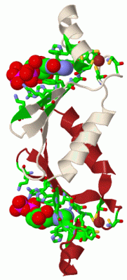
               SCOP domains d1uw1a_ A: Artificial nucleotide binding protein (ANBP)             SCOP domains
               CATH domains ------------------------------------------------------------------- CATH domains
               Pfam domains ------------------------------------------------------------------- Pfam domains
         Sec.struct. author .hhhhhhhhhhhhh............eeee..eeeee..hhhhhhhhhhhh......eeeee..... Sec.struct. author
                 SAPs(SNPs) ------------------------------------------------------------------- SAPs(SNPs)
                    PROSITE ------------------------------------------------------------------- PROSITE
                 Transcript ------------------------------------------------------------------- Transcript
                  1uw1 A  7 DDKKTNWLKRIYRVRPCVKCKVAPRNWKVKNKHLRIYNMCKTCFNNSIDIGDDTYHGHDDWLMYADS 73
                                    16        26        36        46        56        66       

   Legend:   → Mismatch (orange background)
  - → Gap (green background, '-', border residues have a numbering label)
    → Modified Residue (blue background, lower-case, 'x' indicates undefined single-letter code, labelled with number + name)
  x → Chemical Group (purple background, 'x', labelled with number + name, e.g. ACE or NH2)
  extra numbering lines below/above indicate numbering irregularities and modified residue names etc., number ends below/above '|'

 Classification and Annotation

(-) SCOP Domains  (1, 1)

Asymmetric Unit

(-) CATH Domains  (0, 0)

(no "CATH Domain" information available for 1UW1)

(-) Pfam Domains  (0, 0)

(no "Pfam Domain" information available for 1UW1)

(-) Gene Ontology  (0, 0)

Asymmetric Unit(hide GO term definitions)
    (no "Gene Ontology" information available for 1UW1)

 Visualization

(-) Interactive Views

Asymmetric Unit
  Complete Structure
    Jena3D(integrated viewing of ligand, site, SAP, PROSITE, SCOP information)
    WebMol | AstexViewer[tm]@PDBe
(Java Applets, require no local installation except for Java; loading may be slow)
    STRAP
(Java WebStart application, automatic local installation, requires Java; full application with system access!)
    RasMol
(require local installation)
    Molscript (VRML)
(requires installation of a VRML viewer; select preferred view via VRML and generate a mono or stereo PDF format file)
 
  Ligands, Modified Residues, Ions
    ADP  [ RasMol | Jena3D ]  +environment [ RasMol | Jena3D ]
    ZN  [ RasMol | Jena3D ]  +environment [ RasMol | Jena3D ]
 
  Sites
    AC1  [ RasMol ]  +environment [ RasMol ]
    AC2  [ RasMol ]  +environment [ RasMol ]
 
  Cis Peptide Bonds
(no "Cis Peptide Bonds" information available for 1uw1)
 
Biological Unit
  Complete Structure
    Biological Unit 1  [ Jena3D ]

(-) Still Images

Jmol
  protein: cartoon or spacefill or dots and stick; nucleic acid: cartoon and stick; ligands: spacefill; active site: stick
Molscript
  protein, nucleic acid: cartoon; ligands: spacefill; active site: ball and stick

 Databases and Analysis Tools

(-) Databases

Access by PDB/NDB ID
  1uw1
    Family and Domain InformationProDom | SYSTERS
    General Structural InformationGlycoscienceDB | MMDB | NDB | OCA | PDB | PDBe | PDBj | PDBsum | PDBWiki | PQS | PROTEOPEDIA
    Orientation in MembranesOPM
    Protein SurfaceSURFACE
    Secondary StructureDSSP (structure derived) | HSSP (homology derived)
    Structural GenomicsGeneCensus
    Structural NeighboursCE | VAST
    Structure ClassificationCATH | Dali | SCOP
    Validation and Original DataBMRB Data View | BMRB Restraints Grid | EDS | PROCHECK | RECOORD | WHAT_CHECK
 
Access by UniProt ID/Accession number
  (no 'UniProt ID/Accession number' available) |
    Comparative Protein Structure ModelsModBase
    Genomic InformationEnsembl
    Protein-protein InteractionDIP
    Sequence, Family and Domain InformationInterPro | Pfam | SMART | UniProt
 
Access by Enzyme Classificator   (EC Number)
  (no 'Enzyme Classificator' available)
    General Enzyme InformationBRENDA | EC-PDB | Enzyme | IntEnz
    PathwayKEGG | MetaCyc
 
Access by Disease Identifier   (MIM ID)
  (no 'MIM ID' available)
    Disease InformationOMIM
 
Access by GenAge ID
  (no 'GenAge ID' available)
    Age Related InformationGenAge

(-) Analysis Tools

Access by PDB/NDB ID
    Domain InformationXDom
    Interatomic Contacts of Structural UnitsCSU
    Ligand-protein ContactsLPC
    Protein CavitiescastP
    Sequence and Secondary StructurePDBCartoon
    Structure AlignmentSTRAP(Java WebStart application, automatic local installation, requires Java; full application with system access!)
    Structure and Sequence BrowserSTING
 
Access by UniProt ID/Accession number
  (no 'UniProt ID/Accession number' available) |
    Protein Disorder PredictionDisEMBL | FoldIndex | GLOBPLOT

 Related Entries

(-) Entries Sharing at Least One Protein Chain (UniProt ID)

(no "Entries Sharing at Least One Protein Chain" available for 1UW1)

(-) Related Entries Specified in the PDB File

(no "Related Entries Specified in the PDB File" available for 1UW1)