Show PDB file:   
         Plain Text   HTML   (compressed file size)
QuickSearch:   
by PDB,NDB,UniProt,PROSITE Code or Search Term(s)  
(-)Asym./Biol. Unit
(-)Asym./Biol. Unit - sites
collapse expand < >
Image Asym./Biol. Unit
Asym./Biol. Unit  (Jmol Viewer)
Image Asym./Biol. Unit - sites
Asym./Biol. Unit - sites  (Jmol Viewer)

(-) Description

Title :  THREE-DIMENSIONAL CRYSTAL STRUCTURE OF DISSIMILATORY SULFITE REDUCTASE D (DSRD)
 
Authors :  N. Mizuno, G. Voordouw, K. Miki, A. Sarai, Y. Higuchi
Date :  18 Apr 03  (Deposition) - 14 Oct 03  (Release) - 24 Feb 09  (Revision)
Method :  X-RAY DIFFRACTION
Resolution :  1.20
Chains :  Asym./Biol. Unit :  A,B
Keywords :  Crystal Structure, Dissimilatory Sulfite Reductase D, Dna Binding Motif, Sulfate-Reducing Bacteria, Winged-Helix Motif, Unknown Function (Keyword Search: [Gene Ontology, PubMed, Web (Google))
 
Reference :  N. Mizuno, G. Voordouw, K. Miki, A. Sarai, Y. Higuchi
Crystal Structure Of Dissimilatory Sulfite Reductase D (Dsrd) Protein-Possible Interaction With B- And Z-Dna By Its Winged-Helix Motif
Structure V. 11 1133 2003
PubMed-ID: 12962631  |  Reference-DOI: 10.1016/S0969-2126(03)00156-4
(for further references see the PDB file header)

(-) Compounds

Molecule 1 - PROTEIN DSVD
    ChainsA, B
    EngineeredYES
    Expression SystemESCHERICHIA COLI
    Expression System Taxid562
    Organism ScientificDESULFOVIBRIO VULGARIS
    Organism Taxid881
    SynonymDISSIMILATORY SULFITE REDUCTASE D, DSRD

 Structural Features

(-) Chains, Units

  12
Asymmetric/Biological Unit AB

Summary Information (see also Sequences/Alignments below)

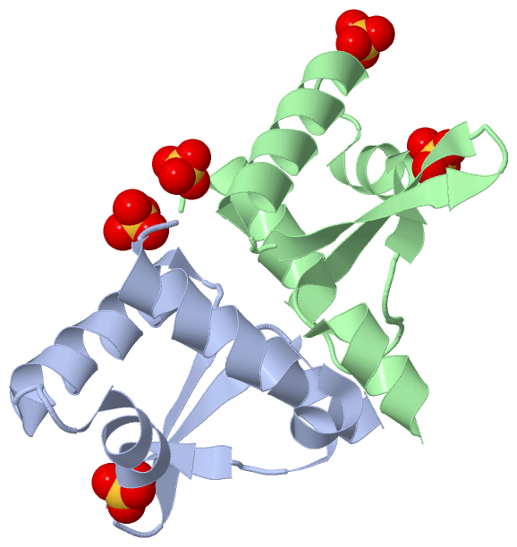
(-) Ligands, Modified Residues, Ions  (1, 5)

Asymmetric/Biological Unit (1, 5)
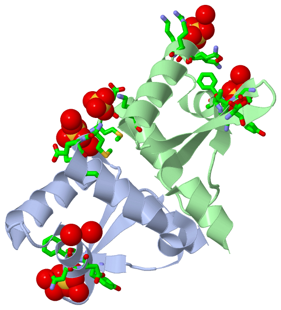
No.NameCountTypeFull Name
1SO45Ligand/IonSULFATE ION

(-) Sites  (5, 5)

Asymmetric Unit (5, 5)
No.NameEvidenceResiduesDescription
1AC1SOFTWAREHOH A:335 , HOH A:389 , LYS B:35 , GLN B:36 , ARG B:37 , HOH B:418BINDING SITE FOR RESIDUE SO4 B 201
2AC2SOFTWAREMET A:1 , GLU A:2 , LYS A:5 , HOH A:383 , HOH A:422BINDING SITE FOR RESIDUE SO4 A 202
3AC3SOFTWAREMET B:1 , GLU B:2 , LYS B:5BINDING SITE FOR RESIDUE SO4 B 203
4AC4SOFTWARESER A:56 , TYR B:23 , PHE B:24 , ASN B:25 , THR B:60 , THR B:61 , HOH B:456BINDING SITE FOR RESIDUE SO4 B 204
5AC5SOFTWARETYR A:23 , PHE A:24 , ASN A:25 , ASP A:26 , THR A:60 , THR A:61 , HOH A:322 , HOH A:331 , HOH A:375 , ASP B:29 , SER B:56BINDING SITE FOR RESIDUE SO4 A 205

(-) SS Bonds  (0, 0)

(no "SS Bond" information available for 1UCR)

(-) Cis Peptide Bonds  (0, 0)

(no "Cis Peptide Bond" information available for 1UCR)

 Sequence-Structure Mapping

(-) SAPs(SNPs)/Variants  (0, 0)

(no "SAP(SNP)/Variant" information available for 1UCR)

(-) PROSITE Motifs  (0, 0)

(no "PROSITE Motif" information available for 1UCR)

(-) Exons   (0, 0)

(no "Exon" information available for 1UCR)

(-) Sequences/Alignments

Asymmetric/Biological Unit
   Reformat: Number of residues per line =  ('0' or empty: single-line sequence representation)
  Number of residues per labelling interval =   
  UniProt sequence: complete  aligned part    
   Show mapping: SCOP domains CATH domains Pfam domains Secondary structure (by author)
SAPs(SNPs) PROSITE motifs Exons
(details for a mapped element are shown in a popup box when the mouse pointer rests over it)
Chain A from PDB  Type:PROTEIN  Length:74
 aligned with DSVD_DESVH | Q46582 from UniProtKB/Swiss-Prot  Length:78

    Alignment length:74
                                    10        20        30        40        50        60        70    
            DSVD_DESVH    1 MEEAKQKVVDFLNSKSGSKSKFYFNDFTDLFPDMKQREVKKILTALVNDEVLEYWSSGSTTMYGLKGAGKQAAA 74
               SCOP domains d1ucra_ A: Dissimilatory sulfite reductase DsvD                            SCOP domains
               CATH domains 1ucrA00 A:1-74 'winged helix' repressor DNA binding domain                 CATH domains
               Pfam domains -------------------------------------------------------------------------- Pfam domains
         Sec.struct. author .hhhhhhhhhhhhhhhhhh..eehhhhhhhh...hhhhhhhhhhhhhhh..eeeeee..eeeeee.hhhhhhhh Sec.struct. author
                 SAPs(SNPs) -------------------------------------------------------------------------- SAPs(SNPs)
                    PROSITE -------------------------------------------------------------------------- PROSITE
                 Transcript -------------------------------------------------------------------------- Transcript
                  1ucr A  1 MEEAKQKVVDFLNSKSGSKSKFYFNDFTDLFPDMKQREVKKILTALVNDEVLEYWSSGSTTMYGLKGAGKQAAA 74
                                    10        20        30        40        50        60        70    

Chain B from PDB  Type:PROTEIN  Length:75
 aligned with DSVD_DESVH | Q46582 from UniProtKB/Swiss-Prot  Length:78

    Alignment length:75
                                    10        20        30        40        50        60        70     
            DSVD_DESVH    1 MEEAKQKVVDFLNSKSGSKSKFYFNDFTDLFPDMKQREVKKILTALVNDEVLEYWSSGSTTMYGLKGAGKQAAAE 75
               SCOP domains d1ucrb_ B: Dissimilatory sulfite reductase DsvD                             SCOP domains
               CATH domains 1ucrB00 B:1-75 'winged helix' repressor DNA binding domain                  CATH domains
           Pfam domains (1) --DsrD-1ucrB01 B:3-69                                                ------ Pfam domains (1)
           Pfam domains (2) --DsrD-1ucrB02 B:3-69                                                ------ Pfam domains (2)
         Sec.struct. author .hhhhhhhhhhhh........eehhhhhhhh...hhhhhhhhhhhhhhh..eeeeee..eeeeee.hhhhhhhhh Sec.struct. author
                 SAPs(SNPs) --------------------------------------------------------------------------- SAPs(SNPs)
                    PROSITE --------------------------------------------------------------------------- PROSITE
                 Transcript --------------------------------------------------------------------------- Transcript
                  1ucr B  1 MEEAKQKVVDFLNSKSGSKSKFYFNDFTDLFPDMKQREVKKILTALVNDEVLEYWSSGSTTMYGLKGAGKQAAAE 75
                                    10        20        30        40        50        60        70     

   Legend:   → Mismatch (orange background)
  - → Gap (green background, '-', border residues have a numbering label)
    → Modified Residue (blue background, lower-case, 'x' indicates undefined single-letter code, labelled with number + name)
  x → Chemical Group (purple background, 'x', labelled with number + name, e.g. ACE or NH2)
  extra numbering lines below/above indicate numbering irregularities and modified residue names etc., number ends below/above '|'

 Classification and Annotation

(-) SCOP Domains  (1, 2)

Asymmetric/Biological Unit

(-) CATH Domains  (1, 2)

Asymmetric/Biological Unit

(-) Pfam Domains  (1, 2)

Asymmetric/Biological Unit

(-) Gene Ontology  (0, 0)

Asymmetric/Biological Unit(hide GO term definitions)
    (no "Gene Ontology" information available for 1UCR)

 Visualization

(-) Interactive Views

Asymmetric/Biological Unit
  Complete Structure
    Jena3D(integrated viewing of ligand, site, SAP, PROSITE, SCOP information)
    WebMol | AstexViewer[tm]@PDBe
(Java Applets, require no local installation except for Java; loading may be slow)
    STRAP
(Java WebStart application, automatic local installation, requires Java; full application with system access!)
    RasMol
(require local installation)
    Molscript (VRML)
(requires installation of a VRML viewer; select preferred view via VRML and generate a mono or stereo PDF format file)
 
  Ligands, Modified Residues, Ions
    SO4  [ RasMol | Jena3D ]  +environment [ RasMol | Jena3D ]
 
  Sites
    AC1  [ RasMol ]  +environment [ RasMol ]
    AC2  [ RasMol ]  +environment [ RasMol ]
    AC3  [ RasMol ]  +environment [ RasMol ]
    AC4  [ RasMol ]  +environment [ RasMol ]
    AC5  [ RasMol ]  +environment [ RasMol ]
 
  Cis Peptide Bonds
(no "Cis Peptide Bonds" information available for 1ucr)
 

(-) Still Images

Jmol
  protein: cartoon or spacefill or dots and stick; nucleic acid: cartoon and stick; ligands: spacefill; active site: stick
Molscript
  protein, nucleic acid: cartoon; ligands: spacefill; active site: ball and stick

 Databases and Analysis Tools

(-) Databases

Access by PDB/NDB ID
  1ucr
    Family and Domain InformationProDom | SYSTERS
    General Structural InformationGlycoscienceDB | MMDB | NDB | OCA | PDB | PDBe | PDBj | PDBsum | PDBWiki | PQS | PROTEOPEDIA
    Orientation in MembranesOPM
    Protein SurfaceSURFACE
    Secondary StructureDSSP (structure derived) | HSSP (homology derived)
    Structural GenomicsGeneCensus
    Structural NeighboursCE | VAST
    Structure ClassificationCATH | Dali | SCOP
    Validation and Original DataBMRB Data View | BMRB Restraints Grid | EDS | PROCHECK | RECOORD | WHAT_CHECK
 
Access by UniProt ID/Accession number
  DSVD_DESVH | Q46582
    Comparative Protein Structure ModelsModBase
    Genomic InformationEnsembl
    Protein-protein InteractionDIP
    Sequence, Family and Domain InformationInterPro | Pfam | SMART | UniProtKB/SwissProt
 
Access by Enzyme Classificator   (EC Number)
  (no 'Enzyme Classificator' available)
    General Enzyme InformationBRENDA | EC-PDB | Enzyme | IntEnz
    PathwayKEGG | MetaCyc
 
Access by Disease Identifier   (MIM ID)
  (no 'MIM ID' available)
    Disease InformationOMIM
 
Access by GenAge ID
  (no 'GenAge ID' available)
    Age Related InformationGenAge

(-) Analysis Tools

Access by PDB/NDB ID
    Domain InformationXDom
    Interatomic Contacts of Structural UnitsCSU
    Ligand-protein ContactsLPC
    Protein CavitiescastP
    Sequence and Secondary StructurePDBCartoon
    Structure AlignmentSTRAP(Java WebStart application, automatic local installation, requires Java; full application with system access!)
    Structure and Sequence BrowserSTING
 
Access by UniProt ID/Accession number
  DSVD_DESVH | Q46582
    Protein Disorder PredictionDisEMBL | FoldIndex | GLOBPLOT (for more information see DisProt)

 Related Entries

(-) Entries Sharing at Least One Protein Chain (UniProt ID)

UniProtKB/Swiss-Prot
        DSVD_DESVH | Q465821wq2

(-) Related Entries Specified in the PDB File

(no "Related Entries Specified in the PDB File" available for 1UCR)