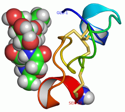
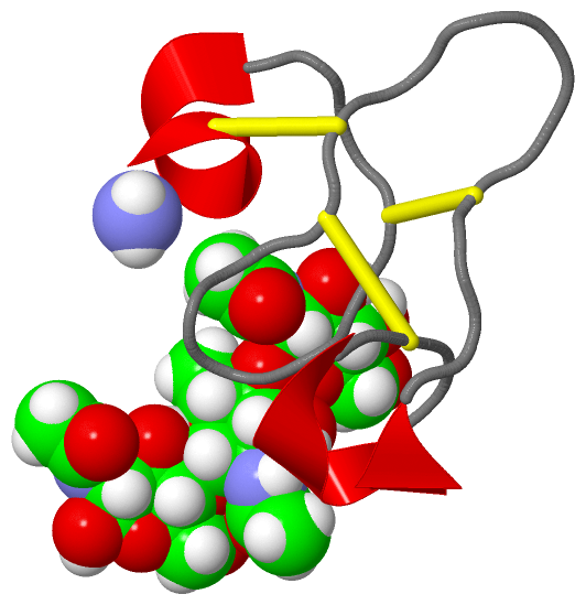
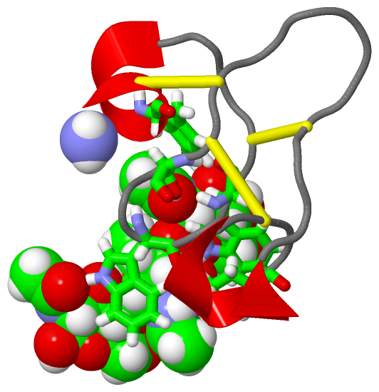
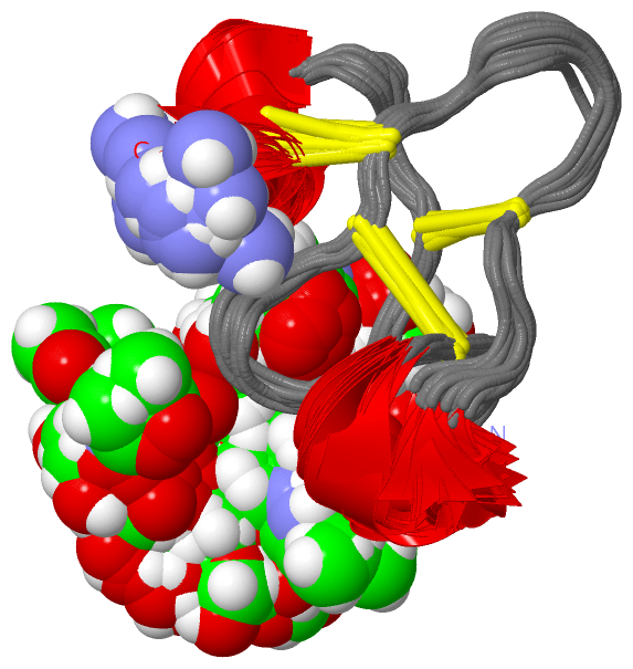
Show PDB file:   
         Plain Text   HTML   (compressed file size)
QuickSearch:   
by PDB,NDB,UniProt,PROSITE Code or Search Term(s)  
(-)NMR Structure - manually
(-)NMR Structure - model 1
(-)NMR Structure - model 1, sites
(-)NMR Structure - all models
collapse expand < >
Image NMR Structure - manually
NMR Structure - manually  (Jmol Viewer)
Image NMR Structure - model 1
NMR Structure - model 1  (Jmol Viewer)
Image NMR Structure - model 1, sites
NMR Structure - model 1, sites  (Jmol Viewer)
Image NMR Structure - all models
NMR Structure - all models  (Jmol Viewer)

(-) Description

Title :  25 NMR STRUCTURES OF TRUNCATED HEVEIN OF 32 AA (HEVEIN-32) COMPLEX WITH N,N,N-TRIACETYLGLUCOSAMINA
 
Authors :  N. Aboitiz, M. Vila-Perello, P. Groves, J. L. Asensio, D. Andreu, F. J. C J. Jimenez-Barbero
Date :  13 Apr 04  (Deposition) - 28 Sep 04  (Release) - 13 Jul 11  (Revision)
Method :  SOLUTION NMR
Resolution :  NOT APPLICABLE
Chains :  NMR Structure  :  A  (25x)
NMR Structure *:  A  (1x)
Keywords :  Alpha-Helix, Anti-Parallel Beta-Sheet, Sugar Binding Protein (Keyword Search: [Gene Ontology, PubMed, Web (Google))
 
Reference :  N. Aboitiz, M. Vila-Perello, P. Groves, J. L. Asensio, D. Andreu, F. J. Canada, J. Jimenez-Barbero
Nmr And Modeling Studies Of Protein-Carbohydrate Interactions: Synthesis, Three-Dimensional Structure, And Recognition Properties Of A Minimum Hevein Domain With Binding Affinity For Chitooligosaccharides
Chembiochem V. 5 1245 2004
PubMed-ID: 15368576  |  Reference-DOI: 10.1002/CBIC.200400025
(for further references see the PDB file header)

(-) Compounds

Molecule 1 - HEVEIN
    ChainsA
    EngineeredYES
    FragmentN-TERMINAL DOMAIN
    Other DetailsSEQUENCE PREPARED ON A MBHA RESIN BY STANDARD SOLID PHASE PEPTIDE SYNTHESIS PROTOCOLS. THE SEQUENCE OF THE PEPTIDE IS NATURALLY FOUND IN HEVEA BRASILIENSIS (PARA RUBBER TREE).
    SynonymALLERGEN HEV B 6
    SyntheticYES

 Structural Features

(-) Chains, Units

  1
NMR Structure (25x)A
NMR Structure * (1x)A

Summary Information (see also Sequences/Alignments below)

(-) Ligands, Modified Residues, Ions  (2, 4)

NMR Structure (2, 4)
No.NameCountTypeFull Name
1NAG3Ligand/IonN-ACETYL-D-GLUCOSAMINE
2NH21Mod. Amino AcidAMINO GROUP
NMR Structure * (2, 4)
No.NameCountTypeFull Name
1NAG3Ligand/IonN-ACETYL-D-GLUCOSAMINE
2NH21Mod. Amino AcidAMINO GROUP

(-) Sites  (3, 3)

NMR Structure (3, 3)
No.NameEvidenceResiduesDescription
1AC1SOFTWARETRP A:21 , NAG A:35BINDING SITE FOR RESIDUE NAG A 34
2AC2SOFTWAREGLU A:1 , TRP A:21 , TRP A:23 , NAG A:34 , NAG A:36BINDING SITE FOR RESIDUE NAG A 35
3AC3SOFTWARESER A:19 , TRP A:23 , TYR A:30 , NAG A:35BINDING SITE FOR RESIDUE NAG A 36

(-) SS Bonds  (3, 3)

NMR Structure
No.Residues
1A:3 -A:18
2A:12 -A:24
3A:17 -A:31

(-) Cis Peptide Bonds  (0, 0)

(no "Cis Peptide Bond" information available for 1T0W)

 Sequence-Structure Mapping

(-) SAPs(SNPs)/Variants  (1, 1)

NMR Structure (1, 1)
  dbSNPPDB
No.SourceVariant IDVariantUniProt IDStatusIDChainVariant
1UniProtVAR_HEVE_HEVBR_001 *N31DHEVE_HEVBR  ---  ---AN14D
   * ID not provided by source

  SNP/SAP Summary Statistics (UniProtKB/Swiss-Prot)
NMR Structure * (1, 1)
  dbSNPPDB
No.SourceVariant IDVariantUniProt IDStatusIDChainVariant
1UniProtVAR_HEVE_HEVBR_001 *N31DHEVE_HEVBR  ---  ---AN14D
   * ID not provided by source

  SNP/SAP Summary Statistics (UniProtKB/Swiss-Prot)

(-) PROSITE Motifs  (1, 1)

NMR Structure (1, 1)
 PROSITEUniProtKBPDB
No.IDACDescriptionIDLocationCountLocation
1CHIT_BIND_I_1PS00026 Chitin recognition or binding domain signature.HEVE_HEVBR29-48  1A:12-31
NMR Structure * (1, 1)
 PROSITEUniProtKBPDB
No.IDACDescriptionIDLocationCountLocation
1CHIT_BIND_I_1PS00026 Chitin recognition or binding domain signature.HEVE_HEVBR29-48  1A:12-31

(-) Exons   (0, 0)

(no "Exon" information available for 1T0W)

(-) Sequences/Alignments

NMR Structure
   Reformat: Number of residues per line =  ('0' or empty: single-line sequence representation)
  Number of residues per labelling interval =   
  UniProt sequence: complete  aligned part    
   Show mapping: SCOP domains CATH domains Pfam domains Secondary structure (by author)
SAPs(SNPs) PROSITE motifs Exons
(details for a mapped element are shown in a popup box when the mouse pointer rests over it)
Chain A from PDB  Type:PROTEIN  Length:33
 aligned with HEVE_HEVBR | P02877 from UniProtKB/Swiss-Prot  Length:204

    Alignment length:33
                                    27        37        47   
            HEVE_HEVBR   18 EQCGRQAGGKLCPNNLCCSQWGWCGSTDEYCSP 50
               SCOP domains d1t0wa_ A: Hevein                 SCOP domains
               CATH domains --------------------------------- CATH domains
               Pfam domains Chitin_bind_1-1t0wA01 A:1-32    - Pfam domains
         Sec.struct. author ...hhhhh..................hhhhhh. Sec.struct. author
                 SAPs(SNPs) -------------D------------------- SAPs(SNPs)
                    PROSITE -----------CHIT_BIND_I_1       -- PROSITE
                 Transcript --------------------------------- Transcript
                  1t0w A  1 EQCGRQAGGKLCPNNLCCSQWGWCGSTDEYCSx 33
                                    10        20        30  |
                                                           33-NH2

   Legend:   → Mismatch (orange background)
  - → Gap (green background, '-', border residues have a numbering label)
    → Modified Residue (blue background, lower-case, 'x' indicates undefined single-letter code, labelled with number + name)
  x → Chemical Group (purple background, 'x', labelled with number + name, e.g. ACE or NH2)
  extra numbering lines below/above indicate numbering irregularities and modified residue names etc., number ends below/above '|'

 Classification and Annotation

(-) SCOP Domains  (1, 1)

NMR Structure

(-) CATH Domains  (0, 0)

(no "CATH Domain" information available for 1T0W)

(-) Pfam Domains  (1, 1)

NMR Structure

(-) Gene Ontology  (3, 3)

NMR Structure(hide GO term definitions)
Chain A   (HEVE_HEVBR | P02877)
molecular function
    GO:0008061    chitin binding    Interacting selectively and non-covalently with chitin, a linear polysaccharide consisting of beta-(1->4)-linked N-acetyl-D-glucosamine residues.
biological process
    GO:0042742    defense response to bacterium    Reactions triggered in response to the presence of a bacterium that act to protect the cell or organism.
    GO:0050832    defense response to fungus    Reactions triggered in response to the presence of a fungus that act to protect the cell or organism.

 Visualization

(-) Interactive Views

NMR Structure
  Complete Structure
    Jena3D(integrated viewing of ligand, site, SAP, PROSITE, SCOP information)
    WebMol | AstexViewer[tm]@PDBe
(Java Applets, require no local installation except for Java; loading may be slow)
    STRAP
(Java WebStart application, automatic local installation, requires Java; full application with system access!)
    RasMol
(require local installation)
    Molscript (VRML)
(requires installation of a VRML viewer; select preferred view via VRML and generate a mono or stereo PDF format file)
 
  Ligands, Modified Residues, Ions
    NAG  [ RasMol | Jena3D ]  +environment [ RasMol | Jena3D ]
    NH2  [ RasMol | Jena3D ]  +environment [ RasMol | Jena3D ]
 
  Sites
    AC1  [ RasMol ]  +environment [ RasMol ]
    AC2  [ RasMol ]  +environment [ RasMol ]
    AC3  [ RasMol ]  +environment [ RasMol ]
 
  Cis Peptide Bonds
(no "Cis Peptide Bonds" information available for 1t0w)
 

(-) Still Images

Jmol
  protein: cartoon or spacefill or dots and stick; nucleic acid: cartoon and stick; ligands: spacefill; active site: stick
Molscript
  protein, nucleic acid: cartoon; ligands: spacefill; active site: ball and stick

 Databases and Analysis Tools

(-) Databases

Access by PDB/NDB ID
  1t0w
    Family and Domain InformationProDom | SYSTERS
    General Structural InformationGlycoscienceDB | MMDB | NDB | OCA | PDB | PDBe | PDBj | PDBsum | PDBWiki | PQS | PROTEOPEDIA
    Orientation in MembranesOPM
    Protein SurfaceSURFACE
    Secondary StructureDSSP (structure derived) | HSSP (homology derived)
    Structural GenomicsGeneCensus
    Structural NeighboursCE | VAST
    Structure ClassificationCATH | Dali | SCOP
    Validation and Original DataBMRB Data View | BMRB Restraints Grid | EDS | PROCHECK | RECOORD | WHAT_CHECK
 
Access by UniProt ID/Accession number
  HEVE_HEVBR | P02877
    Comparative Protein Structure ModelsModBase
    Genomic InformationEnsembl
    Protein-protein InteractionDIP
    Sequence, Family and Domain InformationInterPro | Pfam | SMART | UniProtKB/SwissProt
 
Access by Enzyme Classificator   (EC Number)
  (no 'Enzyme Classificator' available)
    General Enzyme InformationBRENDA | EC-PDB | Enzyme | IntEnz
    PathwayKEGG | MetaCyc
 
Access by Disease Identifier   (MIM ID)
  (no 'MIM ID' available)
    Disease InformationOMIM
 
Access by GenAge ID
  (no 'GenAge ID' available)
    Age Related InformationGenAge

(-) Analysis Tools

Access by PDB/NDB ID
    Domain InformationXDom
    Interatomic Contacts of Structural UnitsCSU
    Ligand-protein ContactsLPC
    Protein CavitiescastP
    Sequence and Secondary StructurePDBCartoon
    Structure AlignmentSTRAP(Java WebStart application, automatic local installation, requires Java; full application with system access!)
    Structure and Sequence BrowserSTING
 
Access by UniProt ID/Accession number
  HEVE_HEVBR | P02877
    Protein Disorder PredictionDisEMBL | FoldIndex | GLOBPLOT (for more information see DisProt)

 Related Entries

(-) Entries Sharing at Least One Protein Chain (UniProt ID)

UniProtKB/Swiss-Prot
        HEVE_HEVBR | P028771hev 1q9b 1wkx 4wp4

(-) Related Entries Specified in the PDB File

1hev NMR STRUCTURE OF HEVEIN (FULL PROTEIN, 43 AA) IN THE FREE STATE
1mmc NMR STRUCTURE OF AC-AMP2 (AMARANTHUS CAUDATUS ANTIMICROBIAL PEPTIDE 2) IN THE FREE STATE RELATED ID: 10261 RELATED DB: BMRB NMR SPECTRAL PARAMETERS OF HEV32 (TRUNCATED HEVEIN OF 32 AA) COMPLEXED WITH CHITOTRIOSE
1q9b CRYSTAL STRUCTURE OF HEVEIN (FULL PROTEIN, 43 AA) IN THE FREE STATE