Show PDB file:   
         Plain Text   HTML   (compressed file size)
QuickSearch:   
by PDB,NDB,UniProt,PROSITE Code or Search Term(s)  
(-)Asymmetric Unit
(-)Asym. Unit - sites
(-)Biological Unit 1
(-)Biol. Unit 1 - sites
(-)Biological Unit 2
collapse expand < >
Image Asymmetric Unit
Asymmetric Unit  (Jmol Viewer)
Image Asym. Unit - sites
Asym. Unit - sites  (Jmol Viewer)
Image Biological Unit 1
Biological Unit 1  (Jmol Viewer)
Image Biol. Unit 1 - sites
Biol. Unit 1 - sites  (Jmol Viewer)
Image Biological Unit 2
Biological Unit 2  (Jmol Viewer)

(-) Description

Title :  STRUCTURE OF AN AAA+ PROTEASE DELIVERY PROTEIN IN COMPLEX WITH A PEPTIDE DEGRADATION TAG
 
Authors :  I. Levchenko, R. A. Grant, D. A. Wah, R. T. Sauer, T. A. Baker
Date :  24 Mar 03  (Deposition) - 23 Sep 03  (Release) - 24 Feb 09  (Revision)
Method :  X-RAY DIFFRACTION
Resolution :  1.60
Chains :  Asym. Unit :  A,B,C,D
Biol. Unit 1:  A (2x),C (1x),D (1x)
Biol. Unit 2:  B  (2x)
Keywords :  Peptide-Binding Pocket, Protein-Peptide Complex, Homodimer, Transport Protein (Keyword Search: [Gene Ontology, PubMed, Web (Google))
 
Reference :  I. Levchenko, R. A. Grant, D. A. Wah, R. T. Sauer, T. A. Baker
Structure Of A Delivery Protein For An Aaa+ Protease In Complex With A Peptide Degradation Tag
Mol. Cell V. 12 365 2003
PubMed-ID: 14536076  |  Reference-DOI: 10.1016/J.MOLCEL.2003.08.014
(for further references see the PDB file header)

(-) Compounds

Molecule 1 - STRINGENT STARVATION PROTEIN B HOMOLOG
    ChainsA, B
    EngineeredYES
    Expression SystemESCHERICHIA COLI
    Expression System Taxid562
    FragmentRESIDUES 1-111
    GeneSSPB
    Organism ScientificHAEMOPHILUS INFLUENZAE
    Organism Taxid727
 
Molecule 2 - SYNTHETIC SSRA PEPTIDE
    ChainsC, D
    EngineeredYES
    Other DetailsTHE PEPTIDE WAS CHEMICALLY SYNTHESIZED.
    SyntheticYES

 Structural Features

(-) Chains, Units

  1234
Asymmetric Unit ABCD
Biological Unit 1 (2x)A (2x) C (1x)D (1x)
Biological Unit 2 (2x) B  

Summary Information (see also Sequences/Alignments below)

(-) Ligands, Modified Residues, Ions  (1, 2)

Asymmetric Unit (1, 2)
No.NameCountTypeFull Name
1MG2Ligand/IonMAGNESIUM ION
Biological Unit 1 (0, 0)
No.NameCountTypeFull Name
1MG-1Ligand/IonMAGNESIUM ION
Biological Unit 2 (0, 0)
No.NameCountTypeFull Name
1MG-1Ligand/IonMAGNESIUM ION

(-) Sites  (2, 2)

Asymmetric Unit (2, 2)
No.NameEvidenceResiduesDescription
1AC1SOFTWARESER A:24 , HOH A:134 , ALA B:56 , THR B:59 , HOH B:169 , HOH B:170BINDING SITE FOR RESIDUE MG B 112
2AC2SOFTWAREALA A:56 , THR A:59 , SER B:24 , HOH B:118BINDING SITE FOR RESIDUE MG A 112

(-) SS Bonds  (0, 0)

(no "SS Bond" information available for 1OU8)

(-) Cis Peptide Bonds  (0, 0)

(no "Cis Peptide Bond" information available for 1OU8)

 Sequence-Structure Mapping

(-) SAPs(SNPs)/Variants  (0, 0)

(no "SAP(SNP)/Variant" information available for 1OU8)

(-) PROSITE Motifs  (0, 0)

(no "PROSITE Motif" information available for 1OU8)

(-) Exons   (0, 0)

(no "Exon" information available for 1OU8)

(-) Sequences/Alignments

Asymmetric Unit
   Reformat: Number of residues per line =  ('0' or empty: single-line sequence representation)
  Number of residues per labelling interval =   
  UniProt sequence: complete  aligned part    
   Show mapping: SCOP domains CATH domains Pfam domains Secondary structure (by author)
SAPs(SNPs) PROSITE motifs Exons
(details for a mapped element are shown in a popup box when the mouse pointer rests over it)
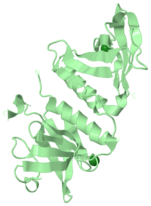
Chain A from PDB  Type:PROTEIN  Length:106
 aligned with SSPB_HAEIN | P45206 from UniProtKB/Swiss-Prot  Length:150

    Alignment length:106
                                    14        24        34        44        54        64        74        84        94       104      
           SSPB_HAEIN     5 SSPKRPYLLRAYYDWLVDNSFTPYLVVDATYLGVNVPVEYVKDGQIVLNLSASATGNLQLTNDFIQFNARFKGVSRELYIPMGAALAIYARENGDGVMFEPEEIYD 110
               SCOP domains d1ou8a_ A: Stringent starvation protein B, SspB                                                            SCOP domains
               CATH domains 1ou8A00 A:5-110 Stringent starvation protein B, SspB                                                       CATH domains
               Pfam domains ---------------------------------------------------------------------------------------------------------- Pfam domains
         Sec.struct. author ...hhhhhhhhhhhhhhhh...eeeeee........hhhhh...eeeee......eeeee...eeeeeeee..eeeeeeee...eeeeee.....eee...hhhhh Sec.struct. author
                 SAPs(SNPs) ---------------------------------------------------------------------------------------------------------- SAPs(SNPs)
                    PROSITE ---------------------------------------------------------------------------------------------------------- PROSITE
                 Transcript ---------------------------------------------------------------------------------------------------------- Transcript
                 1ou8 A   5 SSPKRPYLLRAYYDWLVDNSFTPYLVVDATYLGVNVPVEYVKDGQIVLNLSASATGNLQLTNDFIQFNARFKGVSRELYIPMGAALAIYARENGDGVMFEPEEIYD 110
                                    14        24        34        44        54        64        74        84        94       104      

Chain B from PDB  Type:PROTEIN  Length:106
 aligned with SSPB_HAEIN | P45206 from UniProtKB/Swiss-Prot  Length:150

    Alignment length:106
                                    14        24        34        44        54        64        74        84        94       104      
           SSPB_HAEIN     5 SSPKRPYLLRAYYDWLVDNSFTPYLVVDATYLGVNVPVEYVKDGQIVLNLSASATGNLQLTNDFIQFNARFKGVSRELYIPMGAALAIYARENGDGVMFEPEEIYD 110
               SCOP domains d1ou8b_ B: Stringent starvation protein B, SspB                                                            SCOP domains
               CATH domains 1ou8B00 B:5-110 Stringent starvation protein B, SspB                                                       CATH domains
           Pfam domains (1) SspB-1ou8B01 B:5-110                                                                                       Pfam domains (1)
           Pfam domains (2) SspB-1ou8B02 B:5-110                                                                                       Pfam domains (2)
         Sec.struct. author ...hhhhhhhhhhhhhhhh...eeeeee........hhhhh...eeeee......eeeee...eeeeeeee..eeeeeeee...eeeeee.....eee...hhhhh Sec.struct. author
                 SAPs(SNPs) ---------------------------------------------------------------------------------------------------------- SAPs(SNPs)
                    PROSITE ---------------------------------------------------------------------------------------------------------- PROSITE
                 Transcript ---------------------------------------------------------------------------------------------------------- Transcript
                 1ou8 B   5 SSPKRPYLLRAYYDWLVDNSFTPYLVVDATYLGVNVPVEYVKDGQIVLNLSASATGNLQLTNDFIQFNARFKGVSRELYIPMGAALAIYARENGDGVMFEPEEIYD 110
                                    14        24        34        44        54        64        74        84        94       104      

Chain C from PDB  Type:PROTEIN  Length:11
                                           
               SCOP domains ----------- SCOP domains
               CATH domains ----------- CATH domains
               Pfam domains ----------- Pfam domains
         Sec.struct. author ........... Sec.struct. author
                 SAPs(SNPs) ----------- SAPs(SNPs)
                    PROSITE ----------- PROSITE
                 Transcript ----------- Transcript
                 1ou8 C  97 GRHGAANDENY 107
                                   106 

Chain D from PDB  Type:PROTEIN  Length:8
                                        
               SCOP domains -------- SCOP domains
               CATH domains -------- CATH domains
               Pfam domains -------- Pfam domains
         Sec.struct. author ........ Sec.struct. author
                 SAPs(SNPs) -------- SAPs(SNPs)
                    PROSITE -------- PROSITE
                 Transcript -------- Transcript
                 1ou8 D 100 GAANDENY 107

   Legend:   → Mismatch (orange background)
  - → Gap (green background, '-', border residues have a numbering label)
    → Modified Residue (blue background, lower-case, 'x' indicates undefined single-letter code, labelled with number + name)
  x → Chemical Group (purple background, 'x', labelled with number + name, e.g. ACE or NH2)
  extra numbering lines below/above indicate numbering irregularities and modified residue names etc., number ends below/above '|'

 Classification and Annotation

(-) SCOP Domains  (1, 2)

Asymmetric Unit

(-) CATH Domains  (1, 2)

Asymmetric Unit
(-)
Class: Mainly Beta (13760)

(-) Pfam Domains  (1, 2)

Asymmetric Unit

(-) Gene Ontology  (0, 0)

Asymmetric Unit(hide GO term definitions)
    (no "Gene Ontology" information available for 1OU8)

 Visualization

(-) Interactive Views

Asymmetric Unit
  Complete Structure
    Jena3D(integrated viewing of ligand, site, SAP, PROSITE, SCOP information)
    WebMol | AstexViewer[tm]@PDBe
(Java Applets, require no local installation except for Java; loading may be slow)
    STRAP
(Java WebStart application, automatic local installation, requires Java; full application with system access!)
    RasMol
(require local installation)
    Molscript (VRML)
(requires installation of a VRML viewer; select preferred view via VRML and generate a mono or stereo PDF format file)
 
  Ligands, Modified Residues, Ions
    MG  [ RasMol | Jena3D ]  +environment [ RasMol | Jena3D ]
 
  Sites
    AC1  [ RasMol ]  +environment [ RasMol ]
    AC2  [ RasMol ]  +environment [ RasMol ]
 
  Cis Peptide Bonds
(no "Cis Peptide Bonds" information available for 1ou8)
 
Biological Units
  Complete Structure
    Biological Unit 1  [ Jena3D ]
    Biological Unit 2  [ Jena3D ]

(-) Still Images

Jmol
  protein: cartoon or spacefill or dots and stick; nucleic acid: cartoon and stick; ligands: spacefill; active site: stick
Molscript
  protein, nucleic acid: cartoon; ligands: spacefill; active site: ball and stick

 Databases and Analysis Tools

(-) Databases

Access by PDB/NDB ID
  1ou8
    Family and Domain InformationProDom | SYSTERS
    General Structural InformationGlycoscienceDB | MMDB | NDB | OCA | PDB | PDBe | PDBj | PDBsum | PDBWiki | PQS | PROTEOPEDIA
    Orientation in MembranesOPM
    Protein SurfaceSURFACE
    Secondary StructureDSSP (structure derived) | HSSP (homology derived)
    Structural GenomicsGeneCensus
    Structural NeighboursCE | VAST
    Structure ClassificationCATH | Dali | SCOP
    Validation and Original DataBMRB Data View | BMRB Restraints Grid | EDS | PROCHECK | RECOORD | WHAT_CHECK
 
Access by UniProt ID/Accession number
  SSPB_HAEIN | P45206
    Comparative Protein Structure ModelsModBase
    Genomic InformationEnsembl
    Protein-protein InteractionDIP
    Sequence, Family and Domain InformationInterPro | Pfam | SMART | UniProtKB/SwissProt
 
Access by Enzyme Classificator   (EC Number)
  (no 'Enzyme Classificator' available)
    General Enzyme InformationBRENDA | EC-PDB | Enzyme | IntEnz
    PathwayKEGG | MetaCyc
 
Access by Disease Identifier   (MIM ID)
  (no 'MIM ID' available)
    Disease InformationOMIM
 
Access by GenAge ID
  (no 'GenAge ID' available)
    Age Related InformationGenAge

(-) Analysis Tools

Access by PDB/NDB ID
    Domain InformationXDom
    Interatomic Contacts of Structural UnitsCSU
    Ligand-protein ContactsLPC
    Protein CavitiescastP
    Sequence and Secondary StructurePDBCartoon
    Structure AlignmentSTRAP(Java WebStart application, automatic local installation, requires Java; full application with system access!)
    Structure and Sequence BrowserSTING
 
Access by UniProt ID/Accession number
  SSPB_HAEIN | P45206
    Protein Disorder PredictionDisEMBL | FoldIndex | GLOBPLOT (for more information see DisProt)

 Related Entries

(-) Entries Sharing at Least One Protein Chain (UniProt ID)

UniProtKB/Swiss-Prot
        SSPB_HAEIN | P452061ou9 1oul 1twb 1zsz

(-) Related Entries Specified in the PDB File

1ou9 1oul