Show PDB file:   
         Plain Text   HTML   (compressed file size)
QuickSearch:   
by PDB,NDB,UniProt,PROSITE Code or Search Term(s)  
(-)NMR Structure - model 1
(-)NMR Structure - all models
collapse expand < >
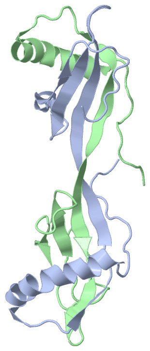
Image NMR Structure - model 1
NMR Structure - model 1  (Jmol Viewer)
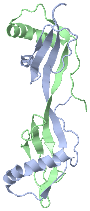
Image NMR Structure - all models
NMR Structure - all models  (Jmol Viewer)

(-) Description

Title :  SOLUTION STRUCTURE OF THE 3D DOMAIN SWAPPED DIMER OF STEFIN A
 
Authors :  R. A. Staniforth, S. Giannini, L. D. Higgins, M. J. Conroy, A. M. Hounslow, R. Jerala, C. J. Craven, J. P. Waltho
Date :  25 Nov 02  (Deposition) - 25 Feb 03  (Release) - 24 Feb 09  (Revision)
Method :  SOLUTION NMR
Resolution :  NOT APPLICABLE
Chains :  NMR Structure  :  A,B
Keywords :  Domain Swapped, Stefin A, Cystatins, Amyloid, Hydrolase Inhibitor (Keyword Search: [Gene Ontology, PubMed, Web (Google)] )
 
Reference :  R. A. Staniforth, S. Giannini, L. D. Higgins, M. J. Conroy, A. M. Hounslow, R. Jerala, C. J. Craven, J. P. Waltho
Three-Dimensional Domain Swapping In The Folded And Molten-Globule States Of Cystatins, An Amyloid-Forming Structural Superfamily
Embo J. V. 20 4774 2001
PubMed-ID: 11532941  |  Reference-DOI: 10.1093/EMBOJ/20.17.4774
(for further references see the PDB file header)

(-) Compounds

Molecule 1 - CYSTATIN A
    Chains: A, B
    Engineered: YES
    Expression System: ESCHERICHIA COLI BL21(DE3)
    Expression System Plasmid: PET3A
    Expression System Strain: BL21-DE3
    Expression System Taxid: 469008
    Expression System Vector Type: PLASMID
    Organism Common: HUMAN
    Organism Scientific: HOMO SAPIENS
    Organism Taxid: 9606
    Synonym: STEFIN A, CYSTATIN AS

 Structural Features

(-) Chains, Units

  
NMR Structure : 

Summary Information (see also Sequences/Alignments below)

(-) Ligands, Modified Residues, Ions  (0, 0)

(no "Ligand,Modified Residues,Ions" information available for 1N9J)

(-) Sites  (0, 0)

(no "Site" information available for 1N9J)

(-) SS Bonds  (0, 0)

(no "SS Bond" information available for 1N9J)

(-) Cis Peptide Bonds  (0, 0)

(no "Cis Peptide Bond" information available for 1N9J)

 Sequence-Structure Mapping

(-) SAPs(SNPs)/Variants  (1, 2)

NMR Structure (1, 2)
  dbSNPPDB
No.SourceVariant IDVariantUniProt IDStatusIDChainVariant
1UniProtVAR_048851T96MCYTA_HUMANPolymorphism34173813A/BT96M

  SNP/SAP Summary Statistics (UniProtKB/Swiss-Prot)

(-) PROSITE Motifs  (1, 2)

NMR Structure (1, 2)
 PROSITEUniProtKBPDB
No.IDACDescriptionIDLocationCountLocation
1CYSTATINPS00287 Cysteine proteases inhibitors signature.CYTA_HUMAN45-58
 
  2A:45-58
B:45-58

(-) Exons   (3, 6)

NMR Structure (3, 6)
 ENSEMBLUniProtKBPDB
No.Transcript IDExonExon IDGenome LocationLengthIDLocationLengthCountLocationLength
1.1aENST000002644741aENSE00001154693chr3:122044091-122044205115CYTA_HUMAN1-22222A:1-22
B:1-22
22
22
1.2aENST000002644742aENSE00001078746chr3:122056394-122056495102CYTA_HUMAN23-56342A:23-56
B:23-56
34
34
1.3ENST000002644743ENSE00001154683chr3:122060286-122060819534CYTA_HUMAN57-98422A:57-98
B:57-98
42
42

(-) Sequences/Alignments

NMR Structure
   Reformat: Number of residues per line =  ('0' or empty: single-line sequence representation)
  Number of residues per labelling interval =   
  UniProt sequence: complete  aligned part    
   Show mapping: SCOP domains CATH domains Pfam domains Secondary structure (by author)
SAPs(SNPs) PROSITE motifs Exons
(details for a mapped element are shown in a popup box when the mouse pointer rests over it)
Chain A from PDB  Type:PROTEIN  Length:98
 aligned with CYTA_HUMAN | P01040 from UniProtKB/Swiss-Prot  Length:98

    Alignment length:98
                                    10        20        30        40        50        60        70        80        90        
            CYTA_HUMAN    1 MIPGGLSEAKPATPEIQEIVDKVKPQLEEKTNETYGKLEAVQYKTQVVAGTNYYIKVRAGDNKYMHLKVFKSLPGQNEDLVLTGYQVDKNKDDELTGF 98
               SCOP domains d1n9ja_ A: Cystatin A (stefin A)                                                                   SCOP domains
               CATH domains 1n9jA00 A:1-98  [code=3.10.450.10, no name defined]                                                CATH domains
               Pfam domains -------------------------------------------------------------------------------------------------- Pfam domains
         Sec.struct. author ............hhhhhhhhhhhhhhhhhhhh....eeee...eeeeeeeeeeeeeee......eeeeeee....hhhhheeeeee............ Sec.struct. author
                 SAPs(SNPs) -----------------------------------------------------------------------------------------------M-- SAPs(SNPs)
                    PROSITE --------------------------------------------CYSTATIN      ---------------------------------------- PROSITE
               Transcript 1 Exon 1.1a  PDB: A:1-22Exon 1.2a  PDB: A:23-56           Exon 1.3  PDB: A:57-98 UniProt: 57-98      Transcript 1
                  1n9j A  1 MIPGGLSEAKPATPEIQEIVDKVKPQLEEKTNETYGKLEAVQYKTQVVAGTNYYIKVRAGDNKYMHLKVFKSLPGQNEDLVLTGYQVDKNKDDELTGF 98
                                    10        20        30        40        50        60        70        80        90        

Chain B from PDB  Type:PROTEIN  Length:98
 aligned with CYTA_HUMAN | P01040 from UniProtKB/Swiss-Prot  Length:98

    Alignment length:98
                                    10        20        30        40        50        60        70        80        90        
            CYTA_HUMAN    1 MIPGGLSEAKPATPEIQEIVDKVKPQLEEKTNETYGKLEAVQYKTQVVAGTNYYIKVRAGDNKYMHLKVFKSLPGQNEDLVLTGYQVDKNKDDELTGF 98
               SCOP domains d1n9jb_ B: Cystatin A (stefin A)                                                                   SCOP domains
               CATH domains 1n9jB00 B:1-98  [code=3.10.450.10, no name defined]                                                CATH domains
           Pfam domains (1) ---Cystatin-1n9jB01 B:4-92                                                                  ------ Pfam domains (1)
           Pfam domains (2) ---Cystatin-1n9jB02 B:4-92                                                                  ------ Pfam domains (2)
         Sec.struct. author ............hhhhhhhhhhhhhhhhhhhh......eeeeeeeeeeeeeeee..eeee.......eeee........eeeee.............. Sec.struct. author
                 SAPs(SNPs) -----------------------------------------------------------------------------------------------M-- SAPs(SNPs)
                    PROSITE --------------------------------------------CYSTATIN      ---------------------------------------- PROSITE
               Transcript 1 Exon 1.1a  PDB: B:1-22Exon 1.2a  PDB: B:23-56           Exon 1.3  PDB: B:57-98 UniProt: 57-98      Transcript 1
                  1n9j B  1 MIPGGLSEAKPATPEIQEIVDKVKPQLEEKTNETYGKLEAVQYKTQVVAGTNYYIKVRAGDNKYMHLKVFKSLPGQNEDLVLTGYQVDKNKDDELTGF 98
                                    10        20        30        40        50        60        70        80        90        

   Legend:   → Mismatch (orange background)
  - → Gap (green background, '-', border residues have a numbering label)
    → Modified Residue (blue background, lower-case, 'x' indicates undefined single-letter code, labelled with number + name)
  x → Chemical Group (purple background, 'x', labelled with number + name, e.g. ACE or NH2)
  extra numbering lines below/above indicate numbering irregularities and modified residue names etc., number ends below/above '|'

 Classification and Annotation

(-) SCOP Domains  (1, 2)

NMR Structure

(-) CATH Domains  (1, 2)

NMR Structure
(-)
Class: Alpha Beta (26913)

(-) Pfam Domains  (1, 2)

NMR Structure
(-)
Clan: Cystatin (20)

(-) Gene Ontology  (19, 19)

NMR Structure(hide GO term definitions)
Chain A,B   (CYTA_HUMAN | P01040)
molecular function
    GO:0004869    cysteine-type endopeptidase inhibitor activity    Stops, prevents or reduces the activity of a cysteine-type endopeptidase, any enzyme that hydrolyzes peptide bonds in polypeptides by a mechanism in which the sulfhydryl group of a cysteine residue at the active center acts as a nucleophile.
    GO:0004866    endopeptidase inhibitor activity    Stops, prevents or reduces the activity of an endopeptidase, any enzyme that hydrolyzes nonterminal peptide bonds in polypeptides.
    GO:0030414    peptidase inhibitor activity    Stops, prevents or reduces the activity of a peptidase, any enzyme that catalyzes the hydrolysis peptide bonds.
    GO:0002020    protease binding    Interacting selectively and non-covalently with any protease or peptidase.
    GO:0030674    protein binding, bridging    The binding activity of a molecule that brings together two or more protein molecules, or a protein and another macromolecule or complex, through a selective, non-covalent, often stoichiometric interaction, permitting those molecules to function in a coordinated way.
    GO:0005198    structural molecule activity    The action of a molecule that contributes to the structural integrity of a complex or its assembly within or outside a cell.
biological process
    GO:0007155    cell adhesion    The attachment of a cell, either to another cell or to an underlying substrate such as the extracellular matrix, via cell adhesion molecules.
    GO:0030216    keratinocyte differentiation    The process in which a relatively unspecialized cell acquires specialized features of a keratinocyte.
    GO:0010951    negative regulation of endopeptidase activity    Any process that decreases the frequency, rate or extent of endopeptidase activity, the endohydrolysis of peptide bonds within proteins.
    GO:0010466    negative regulation of peptidase activity    Any process that stops or reduces the rate of peptidase activity, the hydrolysis of peptide bonds within proteins.
    GO:0045861    negative regulation of proteolysis    Any process that stops, prevents, or reduces the frequency, rate or extent of the hydrolysis of a peptide bond or bonds within a protein.
    GO:0018149    peptide cross-linking    The formation of a covalent cross-link between or within protein chains.
    GO:0016337    single organismal cell-cell adhesion    The attachment of one cell to another cell via adhesion molecules, where both cells are part of the same organism.
cellular component
    GO:0001533    cornified envelope    A type of plasma membrane that has been modified through addition of distinct intracellular and extracellular components, including ceramide, found in cornifying epithelial cells (corneocytes).
    GO:0005737    cytoplasm    All of the contents of a cell excluding the plasma membrane and nucleus, but including other subcellular structures.
    GO:0070062    extracellular exosome    A vesicle that is released into the extracellular region by fusion of the limiting endosomal membrane of a multivesicular body with the plasma membrane. Extracellular exosomes, also simply called exosomes, have a diameter of about 40-100 nm.
    GO:0005615    extracellular space    That part of a multicellular organism outside the cells proper, usually taken to be outside the plasma membranes, and occupied by fluid.
    GO:0005622    intracellular    The living contents of a cell; the matter contained within (but not including) the plasma membrane, usually taken to exclude large vacuoles and masses of secretory or ingested material. In eukaryotes it includes the nucleus and cytoplasm.
    GO:0005634    nucleus    A membrane-bounded organelle of eukaryotic cells in which chromosomes are housed and replicated. In most cells, the nucleus contains all of the cell's chromosomes except the organellar chromosomes, and is the site of RNA synthesis and processing. In some species, or in specialized cell types, RNA metabolism or DNA replication may be absent.

 Visualization

(-) Interactive Views

NMR Structure
  Complete Structure
    Jena3D(integrated viewing of ligand, site, SAP, PROSITE, SCOP information)
    WebMol | AstexViewer[tm]@PDBe
(Java Applets, require no local installation except for Java; loading may be slow)
    STRAP
(Java WebStart application, automatic local installation, requires Java; full application with system access!)
    RasMol
(require local installation)
    Molscript (VRML)
(requires installation of a VRML viewer; select preferred view via VRML and generate a mono or stereo PDF format file)
 
  Ligands, Modified Residues, Ions
(no "Ligands, Modified Residues, Ions" information available for 1n9j)
 
  Sites
(no "Sites" information available for 1n9j)
 
  Cis Peptide Bonds
(no "Cis Peptide Bonds" information available for 1n9j)
 

(-) Still Images

Jmol
  protein: cartoon or spacefill or dots and stick; nucleic acid: cartoon and stick; ligands: spacefill; active site: stick
Molscript
  protein, nucleic acid: cartoon; ligands: spacefill; active site: ball and stick

 Databases and Analysis Tools

(-) Databases

Access by PDB/NDB ID
  1n9j
    Family and Domain Information: ProDom | SYSTERS
    General Structural Information: GlycoscienceDB | MMDB | NDB | OCA | PDB | PDBe | PDBj | PDBsum | PDBWiki | PQS | PROTEOPEDIA
    Orientation in Membranes: OPM
    Protein Surface: SURFACE
    Secondary Structure: DSSP (structure derived) | HSSP (homology derived)
    Structural Genomics: GeneCensus
    Structural Neighbours: CE | VAST
    Structure Classification: CATH | Dali | SCOP
    Validation and Original Data: BMRB Data View | BMRB Restraints Grid | EDS | PROCHECK | RECOORD | WHAT_CHECK
 
Access by UniProt ID/Accession number
  CYTA_HUMAN | P01040
    Comparative Protein Structure Models: ModBase
    Genomic Information: Ensembl
    Protein-protein Interaction: DIP
    Sequence, Family and Domain Information: InterPro | Pfam | SMART | UniProtKB/SwissProt
 
Access by Enzyme Classificator   (EC Number)
  (no 'Enzyme Classificator' available)
    General Enzyme Information: BRENDA | EC-PDB | Enzyme | IntEnz
    Pathway: KEGG | MetaCyc
 
Access by Disease Identifier   (MIM ID)
  (no 'MIM ID' available)
    Disease Information: OMIM
 
Access by GenAge ID
  (no 'GenAge ID' available)
    Age Related Information: GenAge

(-) Analysis Tools

Access by PDB/NDB ID
    Domain Information: XDom
    Interatomic Contacts of Structural Units: CSU
    Ligand-protein Contacts: LPC
    Protein Cavities: castP
    Sequence and Secondary Structure: PDBCartoon
    Structure Alignment: STRAP(Java WebStart application, automatic local installation, requires Java; full application with system access!)
    Structure and Sequence Browser: STING
 
Access by UniProt ID/Accession number
  CYTA_HUMAN | P01040
    Protein Disorder Prediction: DisEMBL | FoldIndex | GLOBPLOT (for more information see DisProt)

 Related Entries

(-) Entries Sharing at Least One Protein Chain (UniProt ID)

UniProtKB/Swiss-Prot
        CYTA_HUMAN | P01040: 1cyu 1cyv 1dvc 1dvd 1gd3 1gd4 1nb3 1nb5 3k9m 3kfq 3kse

(-) Related Entries Specified in the PDB File

1dvc SOLUTION NMR STRUCTURE OF MONOMERIC HUMAN STEFIN A AT PH 5.5 AND 308K